That sinking feeling when unexplained fatigue, kidney stones, or bone pain show up can point to a tiny gland: the parathyroid.
Because the parathyroid and its hormone PTH control blood calcium, small problems can create confusing symptoms and odd lab results.
This guide explains how the parathyroid works, what causes over- and underactivity, and which tests and scans are used.
You’ll learn how primary, secondary, and tertiary hyperparathyroidism differ, what hypoparathyroidism looks like, and the role of vitamin D and kidney disease.
You’ll also discover common diagnostic steps—calcium and PTH testing, sestamibi scans and ultrasound—and when parathyroidectomy may be needed.
By the end, you’ll be able to recognize warning signs and understand treatment choices and recovery expectations.
What does the parathyroid gland do?
The neck contains four tiny glands that sit behind the thyroid. Each gland is about 3 to 5 millimeters across—roughly the size of a grain of rice.
They usually lie on the back of the thyroid lobes but can appear in nearby neck or chest tissue.
Parathyroid tissue secretes parathyroid hormone, often abbreviated PTH. PTH helps regulate blood calcium and keeps serum calcium within a narrow range, roughly 8.6 to 10.2 mg/dL. This tight control matters because calcium affects muscle contraction, nerve signaling, and bone strength.
PTH acts quickly. The hormone has a short half-life, often around 2 to 4 minutes. That short lifespan allows continuous calcium monitoring and minute-by-minute adjustments.
Glands detect small shifts in ionized calcium and signal bones, kidneys, and the gut to release or conserve calcium. Bones can release calcium into the blood. Kidneys can reduce urinary calcium loss and boost active vitamin D production to help gut absorption. (Think of it as a three-way partnership working around the clock.)
Small changes in parathyroid function can affect PTH levels and calcium balance. Overactive glands may raise calcium levels and produce parathyroid disease symptoms. Underactive glands may lower calcium and cause different signs.
It’s important to note that the parathyroid glands are not related to the thyroid gland except by name—”para” means “near.” They have completely different functions, and the thyroid does not regulate calcium.
Authoritative detail is available from the Cleveland Clinic parathyroid gland page.
Not medical advice. Content for informational purposes only. Always consult a qualified healthcare professional for medical advice specific to your situation.
What is parathyroid hormone and how does it work?
Parathyroid hormone (PTH) is a small protein made by chief cells in the four parathyroid glands. The glands sit behind the thyroid in the neck.
Chief cells monitor ionized calcium through the calcium-sensing receptor. Low blood calcium triggers rapid PTH release. High calcium suppresses release. PTH has a short half-life of about 2 to 4 minutes, which allows the body to respond swiftly to calcium changes.
PTH acts on three organs to control calcium. Bone releases calcium when PTH increases osteoclast activity via osteoblast signaling. The process raises blood calcium within minutes.
The kidney responds to PTH by reabsorbing more calcium in distal tubules. The kidney also lowers phosphate reabsorption in proximal tubules, producing phosphaturia. PTH stimulates renal 1-alpha hydroxylase to make active vitamin D.
Active vitamin D raises intestinal calcium absorption. Combined effects on bone, kidney, and gut keep serum calcium within a narrow range.
Normal total serum calcium is roughly 8.6–10.2 mg/dL, and the feedback loop adjusts levels minute to minute. Too much PTH may lead to hyperparathyroidism and high calcium. Too little PTH may lead to hypoparathyroidism and low calcium.
Not medical advice, content for educational purposes only. Always consult a qualified healthcare professional for medical advice specific to your situation.
Understanding hyperparathyroidism and its types
What are the symptoms of hyperparathyroidism?
If your parathyroid glands make too much parathyroid hormone, blood calcium can rise. High calcium levels may affect bones, kidneys, muscles, and the nervous system.
- Parathyroid disease symptoms: bone pain, bone weakening, and a higher risk of fractures.
- Kidney stones occur in many people with high calcium.
- Fatigue and low energy are common complaints.
- Muscle weakness and vague aches may be present.
- Gastrointestinal signs include constipation, nausea, and loss of appetite.
- Mood changes such as depression or memory issues can occur.
Some people have mild or no symptoms despite abnormal PTH levels. Blood tests track PTH and calcium.
Bone turnover may raise alkaline phosphatase; see the page on dangerous alkaline phosphatase level for context.
High calcium levels and abnormal parathyroid hormone levels often drive these signs. Symptom patterns can vary from person to person.
Not medical advice, content for educational purposes only. Always consult a qualified healthcare professional for medical advice specific to your situation.
What is a parathyroid adenoma?
A parathyroid adenoma is a benign parathyroid tumor made of overactive parathyroid cells. It typically affects one gland and prompts excess parathyroid hormone (PTH) release.
Excess PTH raises blood calcium. That can lead to kidney stones, bone loss, fatigue, and muscle weakness.
Adenomas represent about 80% to 85% of cases of primary hyperparathyroidism. Estimates vary, but studies suggest primary hyperparathyroidism appears in roughly 1 to 7 per 1,000 adults.
Some people have enlargement of multiple glands or rare parathyroid cancer. Most adenomas remain benign and are found by blood tests and imaging.
Treatment often targets the overactive gland and aims to normalize PTH and high calcium levels. Outcomes vary by age, gland size, and underlying health.
Not medical advice. Always consult a qualified healthcare professional for medical advice specific to your situation.
What causes secondary hyperparathyroidism?
Secondary hyperparathyroidism occurs when parathyroid glands secrete excess parathyroid hormone to correct low blood calcium.
Vitamin D deficiency lowers calcium absorption from the gut. Low gut absorption triggers higher parathyroid hormone levels. PTH acts on bone, kidney, and intestine to raise calcium.
Chronic kidney disease reduces activation of vitamin D and causes phosphate retention. Low calcium and high phosphate stimulate the glands to produce more PTH. This alters parathyroid gland function and can lead to hyperparathyroidism.
Other contributors include malabsorption, bariatric surgery, and some drugs that block vitamin D or lower calcium. PTH rises often accompany small calcium shifts, which can affect bone strength over months.
Not medical advice, content for educational purposes. Always consult a qualified healthcare professional for medical advice specific to your situation.
What is tertiary hyperparathyroidism?
Tertiary hyperparathyroidism is a rare parathyroid disorder. It follows long-standing secondary hyperparathyroidism that doesn’t resolve.
Parathyroid tissue becomes autonomous and secretes parathyroid hormone (PTH) without normal feedback. This creates overactive parathyroid glands that keep raising calcium levels.
Chronic kidney disease often accompanies the condition. People on long-term dialysis or after kidney transplant may be affected.
Persistent high PTH can raise blood calcium and cause bone loss, muscle weakness, and vascular calcification. Doctors rely on PTH levels, calcium tests, and imaging to confirm the diagnosis.
Treatment options include medical strategies to lower PTH and parathyroid gland surgery. Doctors may perform surgical removal of glands, called parathyroidectomy. Outcomes vary with disease duration and gland changes.
Not medical advice, content for educational purposes. Always consult a qualified healthcare professional for medical advice specific to your situation.
What are the signs of hypoparathyroidism?
Underactive parathyroid glands lower parathyroid hormone (PTH) levels. Low PTH leads to dangerously low calcium in the blood. Symptoms vary from mild to severe and can develop suddenly.
- Tingling or numbness around the mouth and in the fingers.
- Muscle cramps and painful spasms. Severe spasms may affect the throat or breathing muscles.
- Fatigue, anxiety, or memory problems.
- Seizures or fainting in cases of very low calcium.
- Dry skin, brittle nails, and sparse hair over time.
- Heart palpitations related to abnormal heart rhythms.
Surgical injury to the parathyroid glands during thyroid or neck operations is a common cause. Autoimmune disease, genetic conditions, and low magnesium can lead to the same pattern.
Temporary low calcium is reported in up to 20–30% after thyroidectomy. Permanent hypoparathyroidism is reported in about 1–5% of cases.
Diagnosis relies on blood tests showing low calcium with low or inappropriately normal PTH levels. Management is often focused on oral calcium and active vitamin D supplements, with intravenous calcium used for severe symptoms. Long-term follow-up monitors calcium, phosphate, and kidney health to reduce complications.
Parathyroid disease symptoms can vary widely from person to person. Not medical advice, content for educational purposes only. Always consult a qualified healthcare professional for medical advice specific to your situation.
How is primary hyperparathyroidism diagnosed?
Diagnosis begins with blood tests that measure serum calcium and parathyroid hormone levels. Elevated calcium with an elevated or inappropriately normal PTH suggests an overactive parathyroid gland.
- Serum total calcium and ionized calcium.
- PTH assay to compare against calcium.
- Vitamin D and creatinine to check related causes.
- 24-hour urine calcium to assess calcium handling.
- Bone density testing to measure bone loss.
Clinicians interpret PTH relative to calcium. A high calcium value with a PTH above the lab reference supports primary hyperparathyroidism. A normal-range PTH while calcium is high may be inappropriately normal and still indicate disease.
Laboratory cutoffs vary by lab and assay. Results can vary by age and by lab method. For example, a calcium of 11.2 mg/dL with a PTH above the reference range often points toward primary hyperparathyroidism.
Additional tests help define function and risk. Measuring urinary calcium helps separate primary hyperparathyroidism from familial or absorptive causes.
Imaging follows biochemical confirmation. Parathyroid function tests guide surgical planning when needed.
A clear guide on how to read blood test results may help explain common lab values.
Not medical advice, content for educational purposes. Always consult a qualified healthcare professional for medical advice specific to your situation.
Parathyroid imaging and diagnostic scans
What is a parathyroid scan used for?
A sestamibi parathyroid scan helps locate overactive parathyroid glands before surgery. Surgeons use the scan to plan focused parathyroidectomy for primary hyperparathyroidism.
A radioactive tracer, technetium-99m sestamibi, concentrates in mitochondria-rich cells. Overactive parathyroid tissue often shows higher tracer uptake than surrounding neck tissue.
Imaging commonly includes early and delayed views or SPECT/CT to show persistent uptake. Combining functional sestamibi imaging with ultrasound can improve localization.
According to available research, sensitivity ranges 70–90% for single-gland parathyroid adenomas. Sensitivity falls to about 40–60% for multi-gland disease.
Use of SPECT/CT or combined tests raises true-positive rates and reduces operative time. Study results vary by technique, operator, and gland size. Localizing parathyroid adenomas remains less reliable for small or multiple adenomas.
Not medical advice, content for educational purposes. Always consult a qualified healthcare professional for medical advice specific to your situation.
When is parathyroid ultrasound recommended?
Ultrasound visualizes parathyroid glands as small, usually hypoechoic nodules behind the thyroid. It shows gland size and relation to nearby structures.
Ultrasound is non-invasive and uses no radiation. It offers real-time images. These features show parathyroid ultrasound benefits.
Ultrasound misses ectopic glands in the chest and very small adenomas. Its sensitivity varies and depends on operator skill. A sestamibi parathyroid scan can detect deep or ectopic lesions more reliably.
Doctors often order a neck ultrasound when blood tests show high calcium with elevated PTH levels. Ultrasound aids surgical planning and supports focused parathyroid adenoma detection.
Not medical advice, content for educational purposes. Always consult a qualified healthcare professional for medical advice specific to your situation.
How rare is parathyroid cancer?
Parathyroid gland cancer is extremely rare. It accounts for fewer than 1% of primary hyperparathyroidism cases.
Estimates place incidence under one per million people each year. Most parathyroid problems arise from benign adenomas. The parathyroid adenoma prevalence far exceeds cancer.
Clinicians suspect malignancy when high calcium levels appear or parathyroid hormone (PTH) levels rise dramatically. Typical red flags include a palpable neck mass and invasive features on scans.
Blood calcium sometimes exceeds 14 mg/dL, and PTH can reach many times the normal range. Sestamibi parathyroid scan and parathyroid ultrasound may help locate lesions. CT or MRI can show invasion into nearby tissue.
Pathology after parathyroidectomy confirms diagnosis, since needle biopsy can risk tumor spread.
Not medical advice, content for educational purposes. Always consult a qualified healthcare professional for medical advice specific to your situation.
You’ll also like: How To Test Cortisol Levels At Home
What treatments are available for parathyroid disorders?
Treatment depends on the type and severity of parathyroid dysfunction.
| Treatment Type | Indication | Expected Outcome |
|---|---|---|
| Parathyroidectomy | Primary hyperparathyroidism with adenoma | Cure rates ~95% in experienced centers |
| Cinacalcet | Mild or secondary hyperparathyroidism | Lowers calcium by affecting PTH activity |
| Vitamin D & phosphate management | Secondary hyperparathyroidism from kidney disease | Helps normalize PTH and calcium levels |
| Active monitoring | Mild, asymptomatic hyperparathyroidism | Tracks calcium and PTH every 6–12 months |
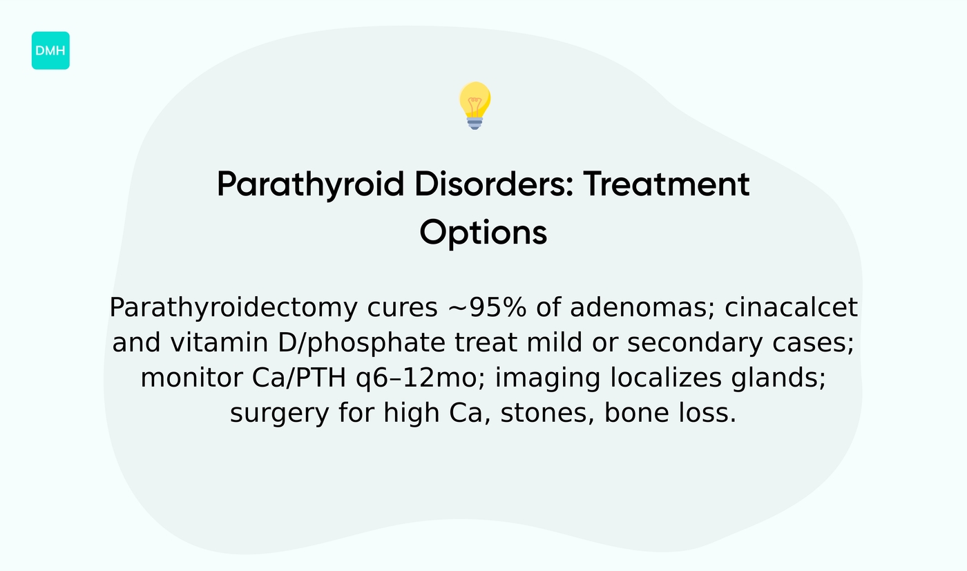
Surgical removal of one or more glands, called a parathyroidectomy, often resolves primary hyperparathyroidism caused by a parathyroid adenoma. Cure rates for a single adenoma can reach about 95% in experienced centers.
Surgery may be recommended for symptomatic high calcium levels, kidney stones, bone loss, or declining kidney function.
Medication can manage mild or secondary cases. Drugs such as cinacalcet may lower calcium by affecting parathyroid hormone activity. Vitamin D and phosphate management can help secondary hyperparathyroidism linked to chronic kidney disease. Bisphosphonates may be used to support bone density in some patients.
Active monitoring suits many people with mild, asymptomatic hyperparathyroidism. Doctors may track serum calcium and PTH levels every 6 to 12 months and assess bone density periodically.
Imaging with a sestamibi parathyroid scan or ultrasound helps localize abnormal glands before surgery. Surgery becomes necessary if symptoms worsen, calcium levels rise, or bone and kidney measures decline. Complex cases such as tertiary hyperparathyroidism may require more extensive gland removal or targeted approaches.
Information about specialist care is available at what kind of doctor to see for parathyroid disease, which outlines common specialist types and evaluations.
Monitor calcium levels and parathyroid hormone levels under medical supervision. Not medical advice, content for educational purposes. Always consult a qualified healthcare professional for medical advice specific to your situation.
Read also: How To Prevent Osteoporosis At Any Age
What is the recovery time after parathyroid surgery?
Recovery after parathyroidectomy varies by case. The parathyroidectomy recovery timeline depends on age, gland size, and overall health.
Most people leave the hospital the same day or after an overnight stay. Some centers report same-day discharge in over 70% of uncomplicated cases.
Neck soreness and mild swelling typically last one to two weeks. Incision pain usually responds to short-term oral pain medication.
Low calcium levels can cause numbness, tingling, or muscle cramps. This transient hypocalcemia risk may occur in up to 30% of patients after surgery.
- Calcium and parathyroid hormone (PTH) levels are checked within 24 hours.
- Ongoing calcium level monitoring often continues at one week and at four to six weeks.
- Some patients need short-term calcium and vitamin D supplements. Supplement needs can vary from a few days to several months.
- Light daily activities often resume within seven days.
- Strenuous exercise and heavy lifting commonly wait three to four weeks.
Long-term outcomes are favorable for most people with primary hyperparathyroidism. Cure rates frequently exceed 95% in experienced centers. Permanent hypoparathyroidism is uncommon and often under 3%.
Not medical advice, content for educational purposes. Always consult a qualified healthcare professional for medical advice specific to your situation.
You might also like: Vitamin D Deficiency: Symptoms, Signs, Causes, and Treatment
Educational notice: This content is provided for informational and educational purposes only and is not intended as medical advice. Always consult a qualified healthcare professional for medical concerns.