Most people treat the thalamus as a simple relay, but it’s actually a dynamic controller tucked above the brainstem.
That narrow view leaves clinicians and families puzzled when thalamic injury causes numbness, altered consciousness, or chronic neuropathic pain.
This guide will unpack thalamus anatomy, major nuclei, thalamocortical pathways, and common disorders so the biology matches the clinical picture.
You’ll learn how the thalamus routes sensory input (except smell), supports attention and sleep-wake states, and shapes higher cognition.
Expect clear, practical explanations and clinical examples that connect findings such as thalamic stroke or hemorrhage to real symptoms and recovery outlook.
By the end you’ll understand why nuclei like the lateral and medial geniculate, pulvinar, and ventral posterior matter for vision, hearing, attention, and touch.
What is the thalamus
The thalamus is a walnut-sized brain structure that sits above the brainstem. It lies near the center of the diencephalon under the cerebral cortex.
Researchers describe the thalamus with a dynamic controller role. It routes most sensory signals to the cortex for conscious perception and shapes cortical rhythms that link attention and consciousness.
The thalamus filters and prioritizes inputs to reduce sensory overload. It coordinates motor signals and supports cognitive processing across networks.
Specialized thalamic nuclei handle distinct tasks:
- The lateral geniculate nucleus processes visual inputs
- The medial geniculate nucleus processes auditory inputs
- The pulvinar nucleus helps visual attention and sensory integration
- The ventral posterior nucleus relays touch, pain, and proprioception
Thalamocortical pathways connect the thalamus with widespread cortical areas and the limbic system. Damage to thalamic tissue may be associated with sensory loss, movement changes, or altered consciousness.
Some studies suggest thalamic injury can affect memory and attention, though effects vary by lesion location and size. Not medical advice; content for educational purposes, consult a professional.
Where is the thalamus located in the brain
The thalamus is a paired, egg-shaped mass of gray matter deep in the brain. It sits in the diencephalon, directly above the brainstem and beneath the cerebral cortex.
The two thalamic bodies lie on either side of the third ventricle near the midline. White matter tracts, including internal capsule fibers, run lateral to each thalamus and connect it to cortical areas.
The thalamus contains about 50 distinct nuclei that serve specialized roles. Nuclei form clusters of gray matter organized around sensory, motor, limbic, and cognitive pathways.
Thalamocortical radiations carry signals between thalamic nuclei and the cortex for perception and attention. The hypothalamus lies just below the thalamus and the epithalamus sits posteriorly.
A small endocrine structure, the pineal gland, lies near the posterior midline and sits close to the dorsal thalamus. Positioning within the diencephalon places the thalamus as a central hub linking the brainstem, limbic structures, and cortex.
This description focuses on anatomy and location for educational clarity. Not medical advice; content for educational purposes, consult a professional.
What is the function of the thalamus
How does the thalamus act as a sensory relay
The thalamus routes most sensory signals to the cerebral cortex for perception. Sensory organs send inputs to specific thalamic nuclei—visual input reaches the lateral geniculate nucleus, auditory input reaches the medial geniculate nucleus, and touch, pain, and proprioception arrive at the ventral posterior nucleus.
The thalamus executes the sensory relay function through several mechanisms. The thalamic reticular nucleus gates attention and sleep rhythms, while thalamocortical loop synchronization times signals for conscious perception.
Signal transmission often occurs within about 20 to 50 ms, according to available research. Olfactory signals bypass the thalamus and connect directly to limbic cortex—some conditions alter sensory routing; see research on which sense is most affected by Alzheimer’s disease.
We describe how the thalamus organizes, prioritizes, and forwards sensory data. Not medical advice; content for educational purposes, consult a professional.
Role in consciousness and sleep regulation
The thalamus helps regulate sleep-wake states and supports conscious cortical dynamics. Thalamic neurons shift firing modes to control state dependency—burst firing promotes sleep rhythms and spindles, while tonic firing supports cortical responsiveness during wakefulness.
Research from George Washington University published in the Journal of Neuroscience reveals the thalamus controls the development of state dependency and continuity. Cellular changes in its relay function are critical for maturing background brain activity and normal sleep-waking states.
During early development the thalamus drives sleep-wake maturation. Thalamocortical circuits form and refine over months, and some studies suggest more stable sleep cycles emerge by about three to six months.
Thalamocortical connections gate sensory flow and synchronize wide cortical networks. Interactions with the thalamic reticular nucleus help generate sleep spindles (~11–16 Hz) and coordinate slow waves. These dynamics are associated with changes in conscious level and with the brain’s ability to integrate sensory and internal signals.
Findings may vary by study design and age. Not medical advice; content for educational purposes, consult a professional.
Executive control and abstract thinking
The thalamus helps guide complex decisions and supports abstract thinking. Recent work from Stony Brook University demonstrates the thalamus plays a decisive role in executive control by actively guiding the brain’s most complex decisions, rather than passively transmitting information.
Using primate models and electrophysiology, researchers found higher-order thalamic nuclei can select behavioral rules and dynamically shape activity in the prefrontal cortex. The thalamus essentially acts as a guide that shapes cortical dynamics for cognitive flexibility.
Evidence comes from neural recordings, reversible inactivation, and human imaging. Some studies report thalamic signals that precede shifts in prefrontal firing by tens to hundreds of milliseconds.
Disrupting specific thalamic nuclei often reduces set-shifting accuracy in animal models. Research points to the mediodorsal and pulvinar nuclei as hubs that route limbic and sensory inputs into executive circuits.
This perspective treats the thalamus as a dynamic controller that coordinates cortical networks for abstract reasoning. Findings vary by species and method, and causality remains under study according to available research. Not medical advice; content for educational purposes, consult a professional.
Attention and memory processing
The thalamus helps direct attention and supports episodic memory through specific nucleus connections. The pulvinar nucleus coordinates visual and spatial attention by shaping thalamocortical pathways and linking sensory cortices.
The thalamic reticular nucleus gates incoming signals with inhibitory control, which refines focus during tasks. During wakefulness it’s involved in selective attention, while during sleep it underlies sleep spindles, according to available research.
The anterior thalamic nuclei connect with the hippocampus and mammillary bodies via the mammillothalamic tract. These limbic links help encode and retrieve episodic memories and integrate emotional context with cortical networks.
Lesions in these thalamic regions associate with attention deficits and memory impairment. Functional MRI and electrophysiology studies report increased thalamocortical synchrony during focused attention and during memory encoding. We describe these roles based on current evidence while noting variability across studies. Not medical advice; content for educational purposes, consult a professional.
Major thalamic nuclei and their functions
| Nucleus | Primary Function | Key Connections |
|---|---|---|
| Lateral Geniculate Nucleus (LGN) | Visual signal relay | Retina → Primary visual cortex |
| Medial Geniculate Nucleus (MGN) | Auditory signal relay | Inferior colliculus → Auditory cortex |
| Pulvinar Nucleus | Visual attention & integration | Visual cortex, parietal areas, superior colliculus |
| Ventral Posterior Nucleus (VPN) | Somatosensory relay | Spinal cord → Somatosensory cortex |
| Thalamic Reticular Nucleus (TRN) | Attention gating & sleep spindles | Lateral thalamic shell → Multiple nuclei |
Lateral geniculate nucleus
The lateral geniculate nucleus (LGN) is a thalamic nucleus that processes visual input from the retina and relays that input to the primary visual cortex.
The LGN organizes retinal signals into six main layers in primates. Two layers form the magnocellular stream for motion and coarse detail, while four layers form the parvocellular stream for color and fine detail. A koniocellular pathway adds short-wavelength contrast information.
The LGN shapes contrast, timing, and spatial detail before cortical transmission. Cortical feedback alters LGN output and gates signals via thalamocortical pathways.
Lesions of the LGN may produce visual field loss or diplopia. See causes of sudden temporary double vision for related symptoms.
We present this summary for informational purposes only. Not medical advice; content for educational purposes, consult a professional.
Medial geniculate nucleus
The medial geniculate nucleus (MGN) is the thalamic relay for auditory signals. It receives input from the inferior colliculus and projects to the auditory cortex for sound analysis.
Neurons in the MGN sort frequency, intensity, and timing. This sorting helps the cortex identify pitch, spatial cues, and complex patterns.
You can view the MGN as a gate that prioritizes important sounds. Thalamocortical pathways carry its output to primary and secondary auditory areas.
Typical MGN response latencies are about 10 to 20 ms after sound onset. Some studies suggest MGN responses adapt with learning and attention. As part of the thalamus sensory relay, the MGN links brainstem auditory centers to cortical interpretation. Not medical advice; content for educational purposes, consult a professional.
Pulvinar nucleus
The pulvinar nucleus is a region of the thalamus that supports visual attention, spatial awareness, and sensory integration. It sits in the posterior thalamus and connects with the visual cortex, parietal areas, and the superior colliculus.
These connections let the pulvinar route, filter, and prioritize visual signals. Imaging studies show increased pulvinar activation during attention shifts, and lesion studies report impaired target selection under competing stimuli.
The pulvinar may coordinate signals across cortical areas via thalamocortical pathways. It integrates visual input with limbic and frontal signals to shape decision thresholds and behavioral responses.
Loss of pulvinar function can alter orienting and attention, reducing accuracy on spatial tasks. We present these findings cautiously; evidence varies by method and task. Not medical advice; content for educational purposes, consult a professional.
Ventral posterior nucleus
The ventral posterior nucleus is a thalamic region that relays somatosensory signals to the cortex. It splits into ventral posterolateral (VPL) and ventral posteromedial (VPM) divisions—VPL carries body input, while VPM carries face and oral input.
Inputs arrive via dorsal column–medial lemniscus for touch and proprioception. Spinothalamic tracts bring pain and temperature signals. Targets include the primary somatosensory cortex for conscious sensation.
The nucleus preserves spatial maps so touch location remains precise. Lesions produce contralateral sensory loss and may cause persistent central pain. Some studies report post-thalamic stroke pain in about 5–10% of patients.
We describe the ventral posterior nucleus as a somatosensory relay that times and routes signals to the sensory cortex, shaping perception at millimeter-scale precision. Not medical advice; content for educational purposes, consult a professional.
Thalamic reticular nucleus
The thalamic reticular nucleus (TRN) is a thin shell of GABAergic neurons that wraps the lateral surface of the thalamus and helps gate thalamic output. During wakefulness the nucleus shapes selective attention by inhibiting competing sensory relay cells.
Some studies suggest this inhibition sharpens signal-to-noise ratios so you focus on a single stimulus among many. During non-REM sleep the nucleus generates sleep spindles, rhythmic 12–15 Hz bursts that last about 0.5–2 seconds.
These spindles may support memory consolidation and cortical plasticity, according to electrophysiology research. Neurons in the nucleus fire rhythmic inhibitory volleys that synchronize thalamocortical loops.
Animal work reports reduced spindle density after TRN disruption, and human imaging links altered TRN activity to attention and sleep differences. We describe known mechanisms while noting uncertainty in causal links. Not medical advice; content for educational purposes, consult a professional.
Thalamocortical pathways and brain connectivity
Thalamocortical radiations are bundles of axons that connect the thalamus to the cerebral cortex. The thalamus acts as a dynamic gate for cortical input, modulating sensory and motor signals via excitatory and inhibitory circuits.
Thalamic projections commonly target cortical layer 4 and deeper layers. These reciprocal loops synchronize timing across regions. The thalamus supports distributed cortical network dynamics by coordinating the timing and gain of cortical responses.
Recent work from Stony Brook University highlights an expanded role of the thalamus in coordinating cortical communication. The thalamus isn’t just a relay center but a dynamic controller of distributed adaptive dynamics within and across cortical networks to support ongoing cognitive tasks.
The thalamus gates information flow through selective relay by distinct nuclei. For example, the lateral geniculate nucleus funnels retinal signals to primary visual cortex while the pulvinar links visual areas with attention systems.
Imaging studies report thalamic bursts that time-lock activity across distant cortical areas. Such timing can alter effective connectivity and influence cognition and attention. Anatomical estimates indicate major thalamocortical tracts contain millions of axons in humans, supporting high-bandwidth signaling between subcortex and cortex.
Thalamus and cortex interactions remain active research areas. We describe findings cautiously, since mechanisms vary by species and method. Not medical advice; content for educational purposes, consult a professional.
How is the thalamus different from the hypothalamus
The thalamus and hypothalamus are adjacent diencephalon structures with different roles. The thalamus sits above the hypothalamus near the top of the brainstem, while the hypothalamus lies below the thalamus and above the pituitary gland.
The thalamus contains many specialized nuclei that route signals. The hypothalamus is smaller and links the brain with the endocrine system.
Here’s how they differ:
- Sensory processing: The thalamus acts as a sensory relay for vision, touch, taste, and hearing pathways. Olfactory signals bypass the thalamus and reach limbic regions directly.
- Consciousness: The thalamus helps regulate alertness and supports cortical activity that underlies consciousness.
- Hormonal control: The hypothalamus controls hormone release via the pituitary and manages body temperature, hunger, thirst, and sleep rhythms.
- Autonomic regulation: The hypothalamus exerts autonomic control over heart rate and blood pressure.
Damage to the thalamus may cause sensory loss, altered consciousness, or cognitive changes. Damage to the hypothalamus may cause hormonal imbalances, appetite changes, or disrupted sleep.
Some studies suggest overlapping roles through limbic and thalamocortical pathways, yet the core distinction remains clear. We present this as an evidence-based summary for educational use. Not medical advice; content for educational purposes, consult a professional.
Thalamus damage symptoms and disorders
Damage to the thalamus causes sensory, motor, cognitive, and consciousness changes. Sensory loss often appears as numbness or altered touch on one body side.
Pain perception can change, producing chronic burning or lancinating pain known as thalamic pain syndrome. Temperature sense and proprioception may decline. Some individuals report dizziness or vertigo; see vertigo causes and treatment for related symptoms and management information.
Motor problems can include weakness on the opposite body side, impaired coordination, and movement irregularities. Small thalamic lesions may disrupt motor planning by altering thalamocortical circuits.
Cognitive impairments frequently affect attention, working memory, and decision processes. Patients may show slowed processing and reduced flexibility in problem solving. These deficits reflect disrupted connections between thalamic nuclei and prefrontal cortex.
Consciousness changes range from mild reduced alertness to coma with bilateral damage. The thalamus helps support wakeful cortical activity, so its injury can change sleep-wake regulation.
Studies on traumatic brain injury show injuries to the thalamus create “attractor dynamics,” where the brain enters a repetitive state it can’t easily escape. This limits available brain states and behaviors, while intact thalamocortical connections support the unpredictability associated with consciousness.
Symptoms vary by lesion location, size, and patient factors. Some studies report persistent sensory or pain issues in a subset of cases, while others improve over months. Not medical advice; content for educational purposes, consult a professional.
What is a thalamic stroke
A thalamic stroke is a type of brain infarct that affects the thalamus. The thalamus sits above the brainstem and helps route sensory and motor signals. A stroke occurs when blood flow to thalamic tissue is interrupted by a clot or bleeding.
Ischemic thalamic strokes may account for about 2–4% of all ischemic strokes, though estimates vary by study and population. Common causes include small-vessel disease, artery occlusion, or less often, embolic events. Hemorrhagic events link to high blood pressure and trauma.
Symptoms often affect sensation and movement. Some people report sudden numbness or tingling on one side of the body. Weakness or coordination problems can appear.
Thalamic lesions may cause abnormal sensations such as burning pain, labeled thalamus damage symptoms in clinical descriptions. Vision and eye comfort may change; some patients note headaches behind the eyes when imaging and evaluation occur. Learn more about those headaches behind the eyes.
Consciousness can be affected when paramedian thalamic arteries are involved. People with thalamic injury to the mediodorsal nucleus are less likely to recover the content of consciousness, supporting the view that thalamic input to the cortex enables rich cortical dynamics associated with consciousness.
Recovery of alertness and cognitive function can vary widely. Some individuals show improvement over weeks to months. Outcome depends on lesion size, location, age, and medical care. Findings draw on radiology and stroke literature and may vary across studies. Not medical advice; content for educational purposes, consult a professional.
What causes thalamic pain syndrome
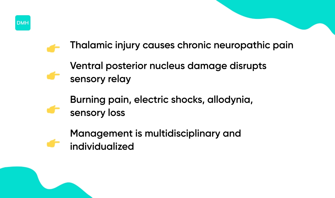
Thalamic pain syndrome describes chronic neuropathic pain after injury to the thalamus. Damage to thalamic nuclei, especially the ventral posterior nucleus, can interrupt normal sensory relay.
This disconnection leads to abnormal signaling in thalamocortical pathways and spinal inputs. Neurons may become hyperexcitable and lose inhibitory control.
Pain usually begins weeks to months after a thalamic stroke or hemorrhage. Estimates vary; some studies report rates from about 2% to 30% among post-stroke cohorts. Onset timing and severity can vary from person to person.
Symptoms commonly include:
- Deep burning pain
- Electric shock sensations
- Abnormal touch perception called allodynia
- Sensory loss coexisting with painful sensations
Triggers often include light touch, temperature shifts, movement, or emotional stress. Small sensory inputs can produce strong pain because central processing becomes amplified.
Underlying mechanisms may include deafferentation (loss of normal input), maladaptive plasticity, central sensitization, and altered thalamic inhibition. Research suggests changes in thalamus limbic system connections may amplify pain perception.
Management is complex and usually multidisciplinary. Evidence about effective treatments varies and depends on individual factors and the exact thalamic lesion. Not medical advice; content for educational purposes, consult a professional.
You might also like: Can Tooth Pain Cause Headaches
Thalamic hemorrhage and treatment approaches
A thalamic hemorrhage is bleeding inside the thalamus, a deep brain relay. High blood pressure often causes thalamic hemorrhage. Head trauma, blood thinners, and vascular malformations can also cause bleeding.
They account for roughly 8–15% of intracerebral hemorrhages in some series. Diagnosis begins with a noncontrast CT scan to detect acute blood quickly. MRI clarifies small bleeds and surrounding tissue injury.
Cerebral angiography identifies aneurysms or arteriovenous malformations when suspected. Head trauma links to post-injury symptoms; see headaches after car accident for related head injury information.
Medical treatment focuses on stabilizing blood pressure and managing intracranial pressure. Targeted reversal of anticoagulants may reduce ongoing bleeding. Neurocritical care teams monitor neurologic status closely.
Surgical options include minimally invasive hematoma evacuation and ventriculostomy to drain cerebrospinal fluid and relieve hydrocephalus. Neurosurgeons select approaches based on hematoma size, location, and patient risk.
Recovery varies by hematoma size, patient age, and treatment speed. Small bleeds often allow partial recovery within months. Large bleeds can cause lasting sensory loss, motor deficits, or thalamic pain syndrome. Rehabilitation commonly includes physical, occupational, and speech therapies.
We present these points for informational purposes only. Not medical advice; content for educational purposes, consult a professional.
You’ll also like: Why Do My Hands Go Numb When I Sleep
The thalamus as a therapeutic target
The thalamus is a central hub that may guide interventions for neurodevelopmental disorders. It gates sensory flow and coordinates cortical rhythms. It links limbic systems and attention networks.
According to researchers at Boston Children’s Hospital, the thalamus is a therapeutic target for neurodevelopmental disorders because it can update its own connections independently of the cortex. Different experiences can change thalamic connectivity in long-lasting ways, making it a strategic focus for treatment.
Some studies suggest thalamic activity shapes learning and cognitive control. Modulating thalamic nodes may influence broad cortical dynamics.
Thalamic neurons can update connections without matching cortical changes. Animal work reports synaptic remodeling in thalamic nuclei after altered sensory experience. Those thalamus connectivity changes can persist for weeks to months and reshape thalamocortical maps. This independent plasticity offers targets distinct from cortical approaches.
Sensory deprivation or enrichment produces lasting thalamic patterns reflecting thalamic adaptive wiring. Rodent studies show increased thalamic synapse density after enrichment. Effects on task performance persist for months.
Human imaging studies probe thalamic involvement in autism and ADHD. According to available research, some reports note changes of roughly 10–30% in functional connectivity metrics, though estimates vary by method. These findings point to a possible neurodevelopmental disorders target for focused research.
Translational and clinical evidence remains limited. Trials that test targeted stimulation or pharmacologic modulation are needed to assess safety and benefit. Not medical advice; content for educational purposes, consult a professional.
You might also like: What Does Sleep Apnea Sound Like
Educational notice: This content is provided for informational and educational purposes only and is not intended as medical advice. Always consult a qualified healthcare professional for medical concerns.