
Most immune tests focus on antibodies, but mononuclear cells do much of the heavy lifting.
That means infections, vaccine responses and immune disorders can be misunderstood when these cells aren’t measured or correctly isolated.
This guide explains how mononuclear cells — monocytes, T and B lymphocytes, and natural killer cells — work and why they matter for both clinical care and research.
It also walks through practical lab steps like PBMC isolation, common markers (CD3, CD14, CD19, CD56), flow cytometry, and best practices for viability and cryopreservation.
You’ll get clear tips to improve PBMC yield and purity (including Ficoll density gradient basics and monocyte isolation methods) so results are more reliable.
By the end, you’ll understand how mononuclear cell counts and functions influence diagnosis, monitoring and experimental outcomes.
What are mononuclear cells
Mononuclear cells refer to white blood cells with a single, round nucleus. These cells include monocytes and lymphocytes, which comprise T cells, B cells, and natural killer cells. Clinicians often call them mononuclear leukocytes.
Peripheral blood mononuclear cells (PBMCs) describes the pool of these cells isolated from blood. PBMC stands for monocytes and lymphocytes in many lab reports.
PBMC isolation methods, such as density gradient centrifugation, separate them from red blood cells and granulocytes. Monocytes often range from about 2% to 8% of circulating white blood cells. Lymphocytes often represent 20% to 40%.
These percentages can vary by age, health, and infection status. Mononuclear cells carry out immune surveillance.
Monocytes may engulf microbes and present antigens. T cells can recognize infected cells and coordinate cellular responses. B cells may produce antibodies that form immune memory. Natural killer cells can kill infected or abnormal cells without prior sensitization.
Researchers use PBMCs to study vaccine responses, infection, and immune activation. Flow cytometry commonly uses markers such as CD3, CD14, CD19, and CD56 to identify subsets.
Some studies suggest PBMC composition may change with disease or treatment, but findings can vary. Not medical advice. For clinical questions about blood tests or cell counts, consult a qualified healthcare professional.
Types of mononuclear cells and their immune functions
Monocytes and their role in immune defense
Monocytes are mononuclear cells in blood. They make about 2–8% of white blood cells. They circulate about one to three days before entering tissues.
In blood they patrol and engulf microbes via phagocytosis. They digest bacteria and clear debris.
They present antigens to other immune cells and trigger broader responses. After entering tissues they differentiate into macrophages and dendritic cells.
Macrophages clear pathogens and dead cells. Dendritic cells specialize in antigen presentation to T cells and help shape adaptive immunity. Monocytes link innate and adaptive systems.
You can find them among peripheral blood mononuclear cells used in PBMC research. Learn more about monocytes for clinical details.
Recent work from Gladstone and UCSF adapted CRISPR-Cas9 gene editing for human monocytes, showing that edited monocytes could still become macrophages and dendritic cells and could still engulf tuberculosis-related microbes in lab tests. Some studies suggest monocyte counts may rise with infection or inflammation.
This content is not medical advice. For medical concerns, consult a qualified healthcare professional.
Lymphocytes including T cells and B cells
Lymphocytes are a type of mononuclear cells. They form a core part of adaptive immunity. They commonly account for about 20–40% of circulating white blood cells.
These cells include T cells and B cells, plus some natural killer cells within PBMC samples. T cells drive T cell responses.
Cytotoxic T cells can kill infected cells. Helper T cells coordinate other immune cells by releasing signaling molecules. B cells make antibodies and can become plasma cells.
Antibodies bind pathogens and mark them for clearance. Memory B cells and T cells may provide long-term immune memory.
Some studies suggest memory cells can last years to decades after exposure. Read more about lymphocytes for detailed clinical information.
For informational purposes only. Not medical advice. Always consult a qualified healthcare professional for personal concerns.
Natural killer cells within PBMCs
Natural killer cells are a subset of lymphocytes found in peripheral blood mononuclear samples. They act as innate immune effectors and don’t require prior antigen sensitization.
These cells identify stressed, virus-infected, or transformed cells. They kill targets by releasing perforin and granzyme proteins. NK cells often make up about 5–15% of lymphocytes recovered from PBMC isolation.
They commonly express CD56 and CD16 surface markers used in PBMC flow cytometry. If you measure cytotoxic activity, NK cells can drive much of the immediate killing seen in assays.
Typical assays include flow-based killing or enzyme-release readouts. NK subsets vary between donors and with age. Some studies suggest that function and frequency can shift after infection or during cancer.
For informational purposes only. Not medical advice.
What are peripheral blood mononuclear cells (PBMCs)
This term refers to mononuclear leukocytes that come from peripheral blood. PBMCs contain a single round nucleus and exclude red blood cells and granulocytes.
Monocytes and lymphocytes make up most PBMCs. Monocytes can perform phagocytosis and may become macrophages or dendritic cells. Lymphocytes include T cells, B cells, and natural killer cells.
Natural killer cells may detect virus-infected or abnormal cells without prior sensitization. Lab teams isolate PBMCs using PBMC isolation methods such as Ficoll density gradient centrifugation.
The Ficoll gradient separates cells by buoyant density. Yields can vary by donor and technique and may range from about 1 to 5 million cells per milliliter of blood. Researchers assess PBMC viability with trypan blue or flow cytometry.
PBMCs often undergo short-term culture for functional assays. Cryopreservation with controlled-rate freezing maintains viability for later use. PBMCs play a central role in immunology research and clinical testing.
Labs use them to measure vaccine responses, profile immune activation, and test cell therapies. Flow cytometry panels targeting CD3, CD14, CD19, and CD56 help identify T cells, monocytes, B cells, and NK cells.
Evidence shows PBMC-based assays may inform disease studies and drug development, but results can vary by protocol and sample quality. For informational purposes only. Not medical advice, consult a professional.
Mononuclear cells vs polymorphonuclear cells
Mononuclear cells have a single round nucleus. They include monocytes, lymphocytes, and natural killer cells. Polymorphonuclear granulocytes have multi-lobed nuclei.
Neutrophils, eosinophils, and basophils belong to this group. Monocytes patrol blood and engulf microbes by phagocytosis. They can become macrophages or dendritic cells to present antigens.
Lymphocytes drive adaptive immunity. T cells handle cellular responses. B cells produce antibodies. Natural killer cells kill infected cells without prior sensitization.
Granulocytes mount rapid innate responses. Neutrophils often form 50–70% of white blood cells. Eosinophils rise with parasites and allergic reactions. Basophils release histamine during allergic responses.
Peripheral blood mononuclear refers to mononuclear leukocytes isolated from blood by density gradient centrifugation. PBMCs are common in research, flow cytometry, and functional assays.
Key lab differences help separate these cells. Granulocytes pellet with red blood cells in Ficoll gradients. Mononuclear cells layer at the buffy coat.
Counts vary by health status and age. Patterns such as low lymphocytes and high neutrophils can prompt further evaluation.
Not medical advice. Content for educational purposes only. Always consult a qualified healthcare professional for medical advice specific to your situation.
How to isolate PBMCs from whole blood
Ficoll density gradient centrifugation method
The Ficoll density gradient separates mononuclear cells by buoyant density. Prepare diluted whole blood and gently layer it over Ficoll.
Centrifuge at about 400 x g for 30 minutes at room temperature without brake. Distinct layers form: plasma, a thin buffy coat with PBMCs, Ficoll, then red cells and granulocytes.
Red blood cells and granulocytes have higher density than the Ficoll solution. Mononuclear cells remain at the plasma–Ficoll interface and are collected as PBMCs. Typical PBMC yield may range from 1 to 5 million cells per mL of blood.
Viability checks use trypan blue or flow cytometry to assess cell quality. Minor granulocyte contamination may occur and extra washes can improve purity.
The density gradient centrifugation step is common in PBMC workflows. This mononuclear cell separation supports downstream PBMC assays. Protocols may vary by lab.
Assessing PBMC viability and yield after isolation
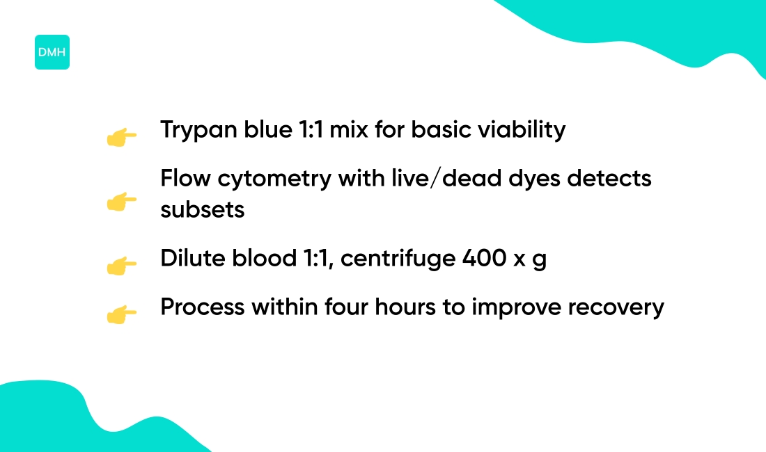
Use simple counts to assess PBMC quality. Mix cells 1:1 with 0.4% trypan blue and count live and dead cells on a hemocytometer.
Trypan blue exclusion often yields viability values above 90% for well-handled samples. Flow cytometry gives a more detailed read.
Use a live/dead dye such as 7-AAD or Zombie plus CD markers to identify monocytes, lymphocytes, and NK cells. PBMC viability testing can reveal subset loss or activation.
Maximize PBMC yield with rapid, gentle processing. Dilute blood 1:1 with PBS, layer carefully over Ficoll, centrifuge at about 400 x g for 30 minutes without brake, then collect the buffy coat.
Wash cells twice at ~300 x g for 10 minutes to reduce platelet carryover. Typical yields range from 0.5–2.0 x 10^6 PBMC per mL of blood, depending on donor and technique.
To improve recovery, process samples within 4 hours and avoid vigorous pipetting. For informational purposes only. Not medical advice, content for educational purposes, consult a professional.
Removing granulocyte contamination from PBMCs
Residual granulocytes can carry over into PBMC preparations and skew results. Use additional wash steps after Ficoll density separation to lower contamination.
Optimizing density gradient centrifugation may reduce granulocyte carryover. Perform two to three washes at 300–400 x g for 8–10 minutes each. Apply ammonium chloride red blood cell lysis for 5–10 minutes at room temperature.
RBC lysis can remove debris and reduce trapped granulocytes. Try 1% dextran sedimentation for about 30 minutes to settle neutrophils. Negative selection with anti-CD15 or CD66b magnetic beads may further deplete granulocytes.
Check purity by flow cytometry using CD3, CD14 and CD15 markers. A target of over 90% mononuclear cells often indicates acceptable purity. High granulocyte levels can mask monocyte and lymphocyte signals.
Small changes in centrifugation speed or time can affect PBMC yield and viability. For informational purposes only. Not medical advice. Consult protocols and vendors.
Common pitfalls in PBMC isolation and how to avoid them
If you isolate PBMCs (peripheral blood mononuclear cells), small errors can cut yield and viability. Typical problems include poor layering, wrong centrifugation, hemolysis, and delayed processing.
Practice a proper layering technique. Slowly dispense diluted blood down the tube wall to form a sharp interface over Ficoll. Avoid mixing the layers during transfer.
Control centrifugation carefully. Use 400–600 x g for 20–30 minutes at room temperature with the brake off. Strong braking or incorrect g values can push granulocytes into the PBMC layer.
Avoid hemolysis by gentle handling and isotonic buffers. Rough pipetting, extreme temperatures, and prolonged storage may cause red cell lysis and lower PBMC viability. Process blood within 2–4 hours of draw.
Some labs report viability drops up to 30% after 24 hours. Aim to process blood promptly to protect cell function. Wash cells twice at 300 x g for 8–10 minutes.
Check PBMC viability with trypan blue; good preparations often show 85–95% viability.
PBMC markers for identifying cell subsets
Researchers use surface markers to sort peripheral blood mononuclear cells. Common markers include CD3, CD14, CD19, and CD56.
CD3 T cells label T lymphocytes. Some studies report CD3+ lymphocytes make up about 60–70% of lymphocytes in PBMC samples. This value can vary by individual and method.
CD14 monocytes mark monocytes, the phagocytic mononuclear leukocytes. CD14+ cells often represent roughly 10–20% of total PBMCs and rise with certain infections or inflammation.
CD19 B cells identify B lymphocytes responsible for antibody responses. CD19+ cells commonly form about 5–15% of PBMC lymphocytes. CD56 detects natural killer cells.
NK cells often range from 5–15% of PBMCs. Some NK subsets co-express CD16 or low levels of CD3. Flow cytometry panels use combinations of these markers to define subsets.
Researchers gate out dead cells and doublets. Multi-color staining helps resolve overlapping populations. Marker percentages depend on PBMC isolation, Ficoll gradient use, instrument settings, and gating strategy.
Validate panels with controls and viability dyes. For informational purposes only. Not medical advice, consult a qualified healthcare professional for clinical interpretation.
Analyzing PBMCs by flow cytometry
Flow cytometry uses fluorescent antibodies to tag cell markers on mononuclear cells. This method distinguishes cell types within peripheral blood mononuclear cells and quantifies relative frequencies.
Common markers include CD3 for T cells, CD19 for B cells, CD14 for monocytes, and CD56 for NK cells. Activation markers such as CD69, HLA-DR, and CD25 can indicate recent stimulation.
Intracellular staining detects cytokines like IFN-γ, IL-2, and TNF-α to assess function. Sample prep often uses about 1 x 10⁶ PBMCs per tube with a 20–30 minute antibody incubation at 4°C.
Viability dyes remove dead cells. Compensation controls and fluorescence-minus-one (FMO) controls improve gate accuracy. Typical data collection ranges from 50,000 to 200,000 events per sample depending on the subset frequency.
Multicolor panels commonly span 8–16 channels to separate subsets and activation states. Antibody titration optimizes signal-to-noise and may cut reagent use by 30–70%.
Functional assays can use CFSE or CellTrace to measure proliferation and CD107a to track degranulation. Some studies suggest intracellular cytokine staining can detect responses near 0.1% of total PBMCs in sensitive assays.
Reported results often vary by donor, stimulus, and processing time. For informational purposes only. Always consult a qualified laboratory specialist for protocol details.
Culturing PBMCs in vitro for research
Researchers use peripheral blood mononuclear cells to model immune responses. These mononuclear cells include monocytes, T cells, B cells and natural killer cells.
Common media include RPMI 1640 with 10% heat-inactivated fetal bovine serum. Incubate at 37°C with 5% CO2 to maintain cell health. Typical seeding density ranges from 0.5 to 2 x 10⁶ cells per mL.
Keep volumes shallow to improve gas exchange. For T cell activation, use anti-CD3/CD28 antibodies or phytohemagglutinin at low microgram per milliliter ranges. Add IL-2 at tens to hundreds of units per mL for expansion when needed.
Monocyte differentiation may use M-CSF or GM-CSF at nanogram per milliliter doses to produce macrophages or dendritic cells over five to seven days. Short stimulations of four to 24 hours suit cytokine assays.
Longer culture times of days to weeks suit proliferation or drug-response studies. Monitor viability by trypan blue or flow cytometry. Aim for viability above 85 to 90 percent after isolation for reliable assays.
Keep sterility and gentle handling to limit activation from processing. Avoid high centrifugation speeds that reduce PBMC yield. Applications include vaccine response assays, drug screening, cytokine profiling and PBMC flow cytometry using markers CD3, CD14, CD19 and CD56.
The following tips may help improve results: optimize media supplements, control cell density, and validate stimulation doses in small tests. PBMC culture conditions support reproducible data.
Peripheral blood mononuclear cells respond variably by donor. Mononuclear leukocytes markers guide subset analysis. For informational purposes only. Not medical advice, consult a professional for clinical questions.
How to isolate monocytes from PBMCs
Monocytes are a subset of mononuclear cells that often make up 5–15% of PBMCs. Two common isolation approaches exist for monocyte enrichment.
The CD14 magnetic bead method uses antibodies against CD14. Label PBMCs with beads, pass them through a magnetic column, wash, and elute the bound monocytes.
Labs often report >90% purity and viability above 90%, although yields depend on blood volume and starting PBMC count. The adherence-based method relies on monocyte attachment to tissue-culture plastic.
Plate PBMCs at 1–2 x 10^6 cells per milliliter, incubate 30–60 minutes at 37°C, then gently remove non-adherent cells. Expect lower purity, often 60–80%, and higher risk of activation with longer adhesion.
Choose based on downstream needs. Use magnetic separation for flow cytometry or RNA work that needs high purity. Use adherence when budgets constrain reagents or when slight activation doesn’t affect results.
Monitor viability with trypan blue or flow cytometry. Check purity using CD14 and CD3/CD19 markers. Keep washes cold and minimize processing time to preserve cell quality.
Track yields per milliliter of blood to compare runs and optimize protocols. Protocol specifics vary by kit and lab. Manufacturer instructions and institutional biosafety rules should guide final steps. For informational purposes only. Not medical advice.
PBMC cryopreservation and thawing protocols
Freezing peripheral blood mononuclear cells (PBMCs) preserves viability for research. Use a cryoprotectant such as 10% DMSO solution in fetal bovine serum or defined cryomedia.
Adjust cell concentration to 5–10 x 106 cells per mL per vial. Cool cells at about -1°C per minute with a programmable freezer or an isopropanol-based freezing container.
A controlled-rate freezing protocol reduces ice crystal damage. Transfer vials to liquid nitrogen for long-term storage. Prefer liquid nitrogen storage in the vapor phase at temperatures below -150°C.
Thaw rapidly in a 37°C water bath until a small ice pellet remains, usually one to two minutes. Dilute thawed PBMCs into ten volumes of pre-warmed culture medium to lower DMSO concentration gradually.
Centrifuge, remove supernatant, and resuspend in fresh medium. Assess viability with trypan blue or flow cytometry. Viability often exceeds 80% after optimal handling.
DMSO can harm cells if left at room temperature. Wash cells within ten minutes of thaw when possible. Some studies suggest slower cooling or inadequate dilution lowers PBMC yield and function.
Record freezing rates, cryoprotectant composition, cell concentration, and storage conditions for reproducibility. Not medical advice. Content for educational purposes, consult a qualified professional for protocol validation.
You might also like: Leukocytosis: definition, causes, symptoms and treatments
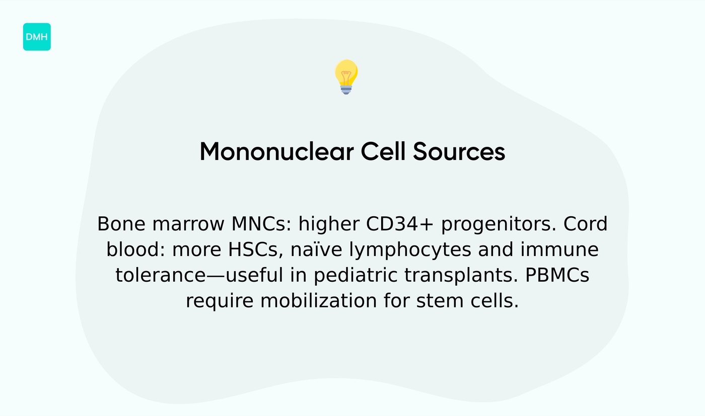
Bone marrow and cord blood mononuclear cells
Bone marrow mononuclear cells contain a mix of progenitors, stromal cells, monocytes, lymphocytes, and hematopoietic stem cells. These cells sit within marrow niches and often show higher proportions of CD34+ progenitors than peripheral blood.
Cord blood mononuclear cells come from the newborn umbilical vein after birth. They carry more naïve lymphocytes and a higher frequency of hematopoietic stem cells per volume compared with adult peripheral blood.
Some studies suggest cord blood shows greater proliferative capacity and immune tolerance. Peripheral blood mononuclear cells (PBMCs) include monocytes, T and B lymphocytes, and natural killer cells.
PBMCs in adults have fewer stem cells unless mobilized with growth factors. Apheresis yields large PBMC collections for transplant or research.
Collection methods differ by source. Bone marrow requires aspiration from the iliac crest under clinical settings. Cord blood is collected at delivery from the clamped cord. Peripheral blood uses venipuncture or apheresis, often after mobilization to increase CD34+ counts.
Clinical use varies by cell source. Bone marrow and mobilized PBMCs often serve in hematopoietic stem cell transplantation for hematologic disorders.
Cord blood units may suit pediatric transplants and some cellular therapies because of HLA tolerance and naïve immune cells. If you review lab samples, expect differences in cell yield, viability, and subset makeup by source.
Evidence shows source selection can affect graft composition and immune reconstitution, but outcomes depend on many clinical factors. For informational purposes only. Always consult a qualified healthcare professional for medical advice specific to your situation.
You might also like: What Are Neutrophils Absolute and Why Do They Matter?
What does it mean when mononuclear cells are high
Mononuclear cells are white blood cells with a single round nucleus. They include monocytes and lymphocytes. A high count means a relative or absolute rise in monocytes or lymphocytes.
Labs report percent and absolute values. Typical monocyte ranges sit at 2–8% (200–800/µL). Lymphocytes often run 20–40% (1,000–4,000/µL). Values above these ranges may prompt follow-up.
Mononuclear cell increase can reflect an immune response. Viral infections such as Epstein-Barr virus and cytomegalovirus often produce lymphocytosis. Chronic infections like tuberculosis and autoimmune conditions such as lupus may raise mononuclear leukocytes.
Certain hematologic disorders can cause sustained elevation. Medication effects and recovery after acute infection may change the PBMC profile. Trends matter more than a single abnormal result.
Clinically concerning signs include persistent fever, night sweats, unexplained weight loss, swollen lymph nodes, and easy bruising. If you’re concerned, should you wait or consult a professional?
Clinicians may order a repeat CBC, peripheral smear, viral serology, or flow cytometry of PBMCs. These tests help distinguish reactive causes from blood disorders.
For specific signs tied to monocyte rises, see high monocyte count symptoms for more detail. Not medical advice. For informational purposes only. Always consult a qualified healthcare professional for medical advice specific to your situation.
Read also: Granulocytes: immature cells, normal range and absolute count
Clinical and research applications of PBMCs
Peripheral blood mononuclear cells are mononuclear cells isolated from blood. They include T cells, B cells, NK cells and monocytes. Labs use them to monitor immune function in patients and studies.
Researchers use PBMC vaccine responses to measure cellular immunity after vaccination. ELISpot assays report spot-forming units per 1,000,000 PBMCs.
Some trials define a response as a fourfold rise or an increase of ~100 spots per 1,000,000 PBMCs. Studies of infectious disease use PBMCs to detect pathogen-specific T cells.
Single-cell RNA sequencing and flow cytometry show functional changes over time. In one Lyme disease study, PBMC profiling helped distinguish Lyme patients from controls and showed increased features linked to T regulatory cells and more monocytes in affected patients.
This approach can help link immune signatures to disease severity. Autoimmunity and cancer research often profile activation markers and cytokine release from PBMCs.
Flow panels using CD3, CD14, CD19 and CD56 quantify subsets. T cells often account for about 50–70% of PBMCs and monocytes about 5–20%. Labs culture PBMCs to test drug effects and antigen responses.
Controlled stimulation can reveal proliferation, cytokine production, or exhaustion markers. Assays may report percent cytokine-positive cells or median fluorescence intensity.
Gene editing research uses PBMCs for ex vivo editing and cell therapy development. Editing can target T cells or other leukocytes to alter function or antigen recognition. PBMC assays offer practical metrics for translational studies and clinical monitoring.
Results vary by protocol and donor. For informational purposes only. Not medical advice, consult a qualified healthcare professional.
You might also like: What Is A Dangerous White Blood Cell Count
Educational notice: This content is provided for informational and educational purposes only and is not intended as medical advice. Always consult a qualified healthcare professional for medical concerns.