Red cell distribution width (RDW) measures how much your red blood cells vary in size.
Labs report it as a percentage, and it shows up on every complete blood count.
A normal RDW means your cells are fairly uniform. A high RDW flags wide size differences — a condition called anisocytosis.
This simple marker gives clinicians early clues about iron shortages, vitamin deficiencies, and other blood disorders. (It’s one of those quiet lab values that packs a real punch.)
Ever wonder why your doctor orders so many follow-up tests after one abnormal CBC? RDW is often the reason — it helps narrow the list of possible causes fast.
What is RDW in a blood test?
Red cell distribution width measures variation in size of red blood cells. Lab machines calculate RDW from red blood cell volume values and plot them on a special graph called a histogram.
If your cells cluster close together on that graph, your RDW stays low. When sizes spread across a wider range, RDW climbs. Labs usually report RDW as a percentage called RDW-CV.
RDW tells you how uniform your red cells are. A low score means similar cell sizes. A high score shows wide size differences.
RDW appears on a complete blood count or CBC report. You can compare RDW with mean corpuscular volume to refine diagnosis. See typical CBC metrics at CBC with differential values and meanings for context.
Well, RDW gives useful early clues about anemia and nutrient shortfalls. Clinicians use RDW with other markers to guide next tests like iron studies and vitamin assays.
If your RDW is abnormal, ask your clinician about iron, vitamin B12, and folate testing. These steps help identify common causes fast.
What is the normal RDW range?
The RDW normal range on most CBC reports reads 11.5% to 14.5%, though some labs may use slightly different cutoffs.
RDW, or red cell distribution width, measures variation in red blood cell size. Labs report RDW two ways: RDW-CV and RDW-SD.
RDW-CV shows as a percentage. RDW-SD shows in femtoliters (fL). Clinical analyzers calculate RDW-CV by dividing the standard deviation of red cell volume by mean corpuscular volume and multiplying by 100.
A normal RDW indicates uniform red cell size and low anisocytosis. You usually see no mixed-size patterns that suggest combined deficiencies when RDW stays inside the normal range.
Actually, RDW is a simple, underused clue for early iron deficiency and other emerging blood issues. If your RDW falls outside 11.5%–14.5%, compare it with MCV and platelet values and ask your clinician about iron studies and vitamin B12 testing.
What does a high RDW mean?
What causes high RDW levels?
High RDW levels signal uneven red blood cell sizes linked to specific medical issues. Doctors recommend testing iron stores, vitamin B12, folate, liver enzymes, and inflammatory markers when RDW climbs above the reference range.
Here’s what commonly drives RDW high causes:
- Iron deficiency anemia — the most common culprit, often raising RDW above 14.5%
- Vitamin B12 or folate deficiency — creates mixed cell sizes as new cells form abnormally
- Liver disease — alters red cell production and shape
- Chronic inflammation or infection — drives variable cell turnover
- Recent transfusion, bone marrow disorders, or hemoglobinopathies such as thalassemia
Common rdw high causes often appear with abnormal MCV or low hemoglobin. Iron problems frequently show an rdw iron deficiency pattern on a CBC.
Checking RDW alongside MCV, ferritin, and B12 speeds diagnosis. If your CBC shows low red cell counts, review this low red blood cell count page and ask your clinician about targeted tests.
What is anisocytosis and how does RDW measure it?
Anisocytosis means your circulating red blood cells differ in volume and shape. Laboratories quantify that variation with RBC distribution width on the CBC.
RDW appears as two measures: RDW-CV given as a percent and RDW-SD given in femtoliters. A high RDW signals mixed cell sizes.
Common causes include iron deficiency, vitamin B12 or folate deficiency, recent bleeding, and some chronic diseases. RDW gives fast, useful clues for diagnosing anemia and guiding follow-up tests like iron studies and B12 levels.
If your RDW is abnormal, review MCV and reticulocyte count and talk with your clinician about next steps. Plus, a peripheral blood smear can show exactly what those size differences look like under the microscope.
When should I be concerned about high RDW?
Be concerned if your RDW sits above the lab reference and you have symptoms. An RDW above 14.5% flags variability in red cell size that needs investigation.
Are you feeling unusually tired or short of breath? Watch for these warning signs:
- Unexplained fatigue, shortness of breath, or fast heartbeat
- New or heavy bleeding, frequent bruising, or dizziness
- Rapid hemoglobin drop or mixed lab patterns like low iron with normal MCV
- Known liver disease, chronic inflammation, or unexplained weight loss
Doctors recommend a prompt repeat CBC with reticulocyte count and targeted studies when symptoms appear. Persistent elevation of red cell distribution width plus symptoms needs timely evaluation.
Ask your clinician for iron studies, B12 and folate levels, and liver tests. Consider hematology referral if results remain abnormal or symptoms worsen.
What does a low RDW indicate?
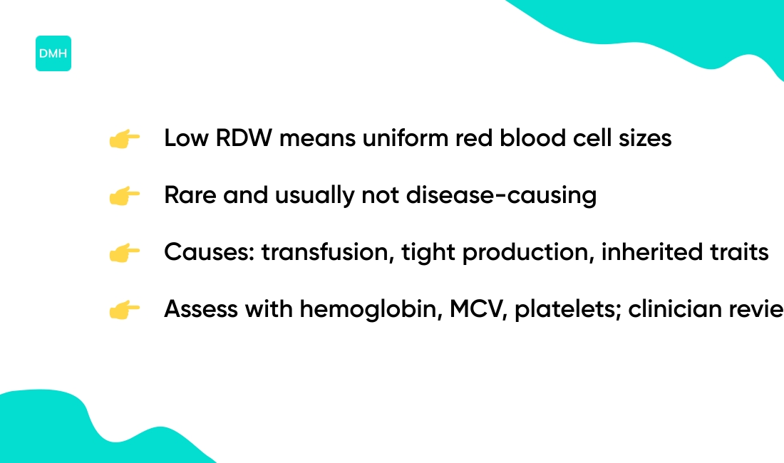
Low RDW usually means your red blood cells are uniform in size. Values below about 11.5% count as low in many labs, though this is quite rare.
Low RDW result rarely points to disease by itself. Common explanations include recent blood transfusion or tight production of similar cells. Some inherited traits produce uniform cells without causing symptoms.
Low RDW doesn’t fit the pattern for iron deficiency or B12 deficiency. Those conditions raise RDW and create anisocytosis. Clinicians interpret RDW with hemoglobin, MCV, and platelet count.
Here’s the thing — a low RDW without other abnormalities usually needs no treatment. Repeating low RDW rarely changes care in clinical practice.
Ask your clinician to review the full CBC and your symptoms for clear guidance. If you want a direct check, request a clinician review of RDW and MCV together.
How do doctors interpret RDW on a CBC?
What is the difference between RDW-CV and RDW-SD?
The two RDW measures used on a CBC serve different purposes. RDW-CV reports the coefficient of variation of red cell volume and shows variation as a percentage. Typical reference sits near 11.5% to 14.5%.
RDW-SD reports the standard deviation of red cell size in femtoliters (fL). Labs often quote values around 39–46 fL.
| Measure | Unit | Normal Range | What It Shows |
|---|---|---|---|
| RDW-CV | Percentage (%) | 11.5–14.5% | Variation relative to mean cell size |
| RDW-SD | Femtoliters (fL) | 39–46 fL | Absolute size spread in cell population |
Use red cell distribution width percent to compare spread against mean cell size. Use RDW-SD to see absolute size spread or mixed populations.
Actually, RDW-SD gives a clearer picture when mean corpuscular volume varies widely. This helps separate iron deficiency from thalassemia. If your rdw blood test is high, review MCV and ask about iron, B12 and folate testing.
How do RDW and MCV together guide diagnosis?
RDW and MCV together classify anemia quickly. RDW measures size variation, while MCV shows average red blood cell size. The pattern they create narrows your differential diagnosis fast.
When RDW rises while MCV falls, expect iron deficiency. When both RDW and MCV rise, suspect vitamin B12 or folate deficiency or mixed causes. When RDW is normal with low MCV, think thalassemia trait.
Here’s a quick reference:
- RDW normal range: 11.5% to 14.5%
- RDW over 14.5% suggests elevation
- MCV under 80 fL indicates microcytosis
- MCV over 100 fL signals macrocytosis
Use RDW-CV and RDW-SD values when available. Pairing RDW with MCV speeds diagnosis and reduces unnecessary tests — research finds RDW adds about 10% diagnostic accuracy when paired with MCV.
Tell your clinician if you have symptoms like fatigue or pallor. Ask them to compare RDW and MCV on your CBC report and explain the pattern they see.
What does elevated RDW with normal MCV mean?
Red cell distribution width above the normal range (about 11.5%–14.5%) with a normal MCV usually means your red cells vary in size while average cell volume stays stable. This pattern flags a mixed or evolving process rather than a single clear anemia type.
Common causes include early iron deficiency, emerging B12 or folate shortage, recent blood loss or transfusion, and a marrow response that raises reticulocytes. (Think of it as catching anemia in transition.)
Your clinician should order these targeted tests:
- An iron studies panel (ferritin, serum iron, TIBC)
- A reticulocyte count test and peripheral smear
- B12 and folate levels, plus hemoglobin electrophoresis if thalassemia trait is suspected
Prioritizing ferritin and a smear guides testing efficiently. Early iron studies identify many cases before MCV changes. See your clinician and ask for these targeted tests to clarify the cause and guide treatment.
What is the relationship between RDW and platelet count?
RDW and platelet indices both signal bone marrow health. The rdw blood test shows size variation of red cells and reflects anisocytosis. Normal RDW sits about 11.5% to 14.5%.
Platelet count and platelet distribution width (PDW) show platelet size and production. PDW can rise with increased platelet production, similar to how RDW rises with variable red cell turnover.
Both reflect bone marrow output and cell turnover. High RDW with abnormal platelets points to marrow stress or mixed deficiencies. For example, iron deficiency often raises RDW and leaves platelets high or even elevated.
Check RDW-CV or RDW-SD when interpreting patterns alongside MCV. If your CBC shows mismatched RDW and platelet values, ask your clinician to request reticulocyte count and iron studies to narrow causes.
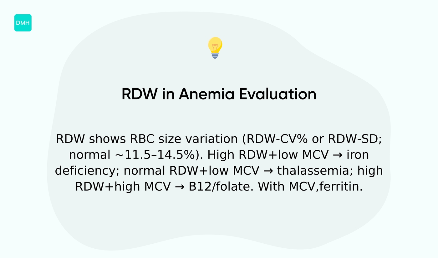
How is RDW used to evaluate anemia?
RDW shows variation in red blood cell size and helps classify anemia quickly. RDW measures anisocytosis and labs report it as RDW-CV percent or RDW-SD in femtoliters.
The RDW blood test helps separate common anemia patterns. A value above the normal range (roughly 11.5% to 14.5%) means cell sizes vary more than expected.
High RDW with low mean corpuscular volume often points toward iron deficiency. Normal RDW with low mean corpuscular volume suggests thalassemia trait. High RDW with high mean corpuscular volume points to vitamin B12 or folate problems.
RDW pairs best with MCV and other markers like reticulocyte count and ferritin. The pattern guides which follow-up test you need and speeds diagnosis. Learn the testing basics on the RDW lab test page.
If you feel fatigue or headaches, check how your RDW fits the full CBC and ask about causes. Read more about anemia symptoms at does anemia cause headaches.
Ask your provider to include RDW when ordering a CBC and to explain the pattern they see. That step often shortens the path to the right treatment.
You’ll also like: Is It Serious To Be Referred To A Hematologist
Can RDW help diagnose iron deficiency?
Rbc distribution width rises early in iron deficiency anemia and flags variation in red cell size. Iron deficiency anemia often shows a widening RDW before hemoglobin falls, giving clinicians an early warning sign.
RDW measures anisocytosis — the mix of small and normal red cells. Iron shortage creates microcytic cells alongside older cells, which widens the distribution.
A high RDW with low mean corpuscular volume points toward iron deficiency rather than thalassemia trait. That pattern helps narrow tests and avoid unnecessary procedures. Values above 14.5% usually raise suspicion.
Clinicians combine RDW with ferritin and transferrin saturation for accuracy. They also check the reticulocyte count to show marrow response.
Actually, many clinicians underuse RDW as an early warning sign. Missed iron deficiency occurs when RDW trends get ignored on routine CBCs.
Use RDW with MCV for clearer interpretation. That approach improves diagnostic accuracy and guides targeted iron studies. If your RDW is high, ask your clinician for iron studies and a reticulocyte count — that’s a practical next step you can take today.
You might also like: What Type Of Cancer Causes Low Hemoglobin
How does RDW help distinguish iron deficiency from thalassemia?
Rbc distribution width reveals variation in red cell size and gives a clear clue when you evaluate anemia. Doctors use RDW with MCV to separate likely iron deficiency from thalassemia trait on initial testing.
Iron deficiency usually shows an rdw high value. Expect RDW above the normal range (about 11.5%–14.5%) and a low MCV under roughly 80 fL. Progressive anisocytosis creates a wider RDW as iron stores fall.
Thalassemia trait often shows a low MCV with a normal RDW. The red cells stay uniformly small, so RDW-CV and RDW-SD remain near normal despite microcytosis. (It’s basically the opposite pattern.)
Use RDW patterns to guide targeted follow-up tests:
- Order ferritin, serum iron, and TIBC to confirm iron deficiency
- Request hemoglobin electrophoresis or HPLC to detect thalassemia trait
- Consider genetic testing when results remain unclear
Combining RDW with MCV and one targeted test will cut diagnostic delay. Learn more about the RDW blood test before you talk with your clinician and ask which follow-up makes sense for your pattern.
Read also: Can Low Iron Cause Headaches