Mean corpuscular volume measures the average size of your red blood cells.
Labs report this number on every complete blood count, and it’s a surprisingly powerful clue about your health.
MCV sits right there on your CBC printout, usually expressed in femtoliters (fL).
When it’s too low or too high, it points your clinician toward specific causes of anemia—or even flags risks you didn’t know existed.
Let’s walk through what MCV really means, how it’s measured, and why you should pay attention to those numbers.
What is MCV in a blood test
Mean corpuscular volume is the average size of your red blood cells. Laboratories report MCV on a complete blood count to show whether your cells are small, normal, or large.
MCV measures volume in femtoliters (fL). Labs calculate it by dividing your hematocrit by your red blood cell count, then multiplying by 1,000.
The MCV on your CBC helps classify anemia as microcytic (small cells), normocytic (normal-sized cells), or macrocytic (large cells). See the CBC with differential values and meanings for related indices like MCH and RDW.
Low MCV points to microcytic anemia from iron deficiency or thalassemia. High MCV points to macrocytic anemia from vitamin B12 or folate deficiency.
I really think MCV gives a quick, useful snapshot of red blood cell health. You should review it alongside other CBC values and targeted tests before accepting a diagnosis.
If your MCV falls outside the lab reference range, ask your clinician about iron studies, B12 testing, folate levels, and a reticulocyte count.
How is MCV calculated and measured
The mean corpuscular volume measures average red blood cell size. Labs report it as femtoliters (fL).
Use the simple formula: MCV (fL) = (Hematocrit % × 10) / RBC (10⁶/µL).
For example, if your hematocrit is 45% and your RBC count is 5.0 million/µL, then MCV = (45 × 10) / 5 = 90 fL. That value falls in a typical adult range.
Automated hematology analyzers provide the raw numbers. Machines count RBCs and determine hematocrit using electrical impedance or optical light scatter to size cells.
The analyzer either calculates MCV from Hct and RBC or derives it directly from the cell volume histogram. Labs flag extreme values for manual review.
Actually, the example makes the concept clear. I’ve seen labs report slightly different MCVs when samples sat too long or when large platelets skew counts.
When you view an MCV on a CBC, note the unit is femtoliters (fL). Ask your clinician about combined indices like MCH and RDW for full interpretation.
What is the normal range for MCV
The standard adult MCV normal range is 80–100 fL. Mean corpuscular volume measures the average red blood cell size on a CBC.
Many laboratories report narrower ranges such as 82–92 fL or 89.8–93.6 fL. Reference ranges vary by analyzer, local population, and lab calibration.
MCV levels shift with age—values tend to rise slightly after age 25. An MCV below about 80 fL suggests microcytic anemia. Common causes include iron deficiency and thalassemia.
An MCV above about 100 fL indicates macrocytic anemia. Causes include vitamin B12 deficiency, folate lack, alcohol use, and liver disease.
A recent Frontiers in Neurology study found a U-shaped association between MCV and stroke outcomes, with an optimal threshold near 92.1 fL. Below that point, each 1 fL increase lowers poor outcome odds by 8%; above it, risk rises 4% per fL.
I’d say reporting your lab’s specific reference range alongside clinical context gives the clearest MCV interpretation. Use MCV with other indices like RDW, MCH, and hemoglobin to clarify causes and guide next tests or treatment.
What does low MCV mean
How iron deficiency affects MCV
Iron shortage shrinks red blood cells and lowers mean corpuscular volume. Low iron limits hemoglobin synthesis, so precursor cells divide more and finish smaller.
Low iron produces microcytic anemia with MCV often under 80 fL. Typical lab patterns show low hemoglobin, low MCV, high RDW, and low ferritin—often below 30 ng/mL.
Iron studies show low serum iron and high TIBC. If you see low MCV with low hemoglobin, check iron tests and look for bleeding or absorption issues. You might also want to read about links with a low red blood cell count.
Treating iron deficiency usually raises MCV within weeks to months, depending on severity and treatment adherence. Talk with your clinician to confirm diagnosis and start targeted therapy.
Other causes of low MCV
Mean corpuscular volume shows average red cell size and guides diagnosis. Low MCV signals microcytic anemia, but not only iron deficiency.
Thalassemia is inherited and causes small red cells with normal iron. Hemoglobin electrophoresis helps confirm the diagnosis.
Chronic blood loss from the gut or heavy menses lowers hemoglobin over time. Stool tests and endoscopy may locate bleeding.
Lead poisoning impairs hemoglobin synthesis. A blood lead test catches this issue early. Sideroblastic anemia and chronic disease can reduce hemoglobin production—ferritin, TIBC, and a bone marrow exam guide workup.
Ordering iron studies plus electrophoresis and a lead level speeds correct diagnosis. Talk to your clinician if your CBC shows low MCV.
Is low MCV always due to iron deficiency
Low MCV isn’t always caused by iron deficiency. A low mean corpuscular volume often signals microcytic anemia, but you need targeted tests to sort causes.
Common causes include iron deficiency, thalassemia, chronic disease, and lead exposure. Lab numbers below 80 fL define microcytosis.
Ferritin and transferrin saturation detect iron deficiency. Ferritin below 30 ng/mL suggests iron deficiency, while hemoglobin electrophoresis identifies thalassemia traits. CRP or ESR helps spot chronic inflammation.
Have you asked your doctor which test comes next? Early testing prevents misdiagnosis. Don’t assume iron is the only culprit.
Start with ferritin, transferrin saturation, and hemoglobin electrophoresis. Follow results with targeted treatment—correct diagnosis guides specific therapy and monitoring.
What does high MCV indicate
Does vitamin B12 deficiency increase MCV
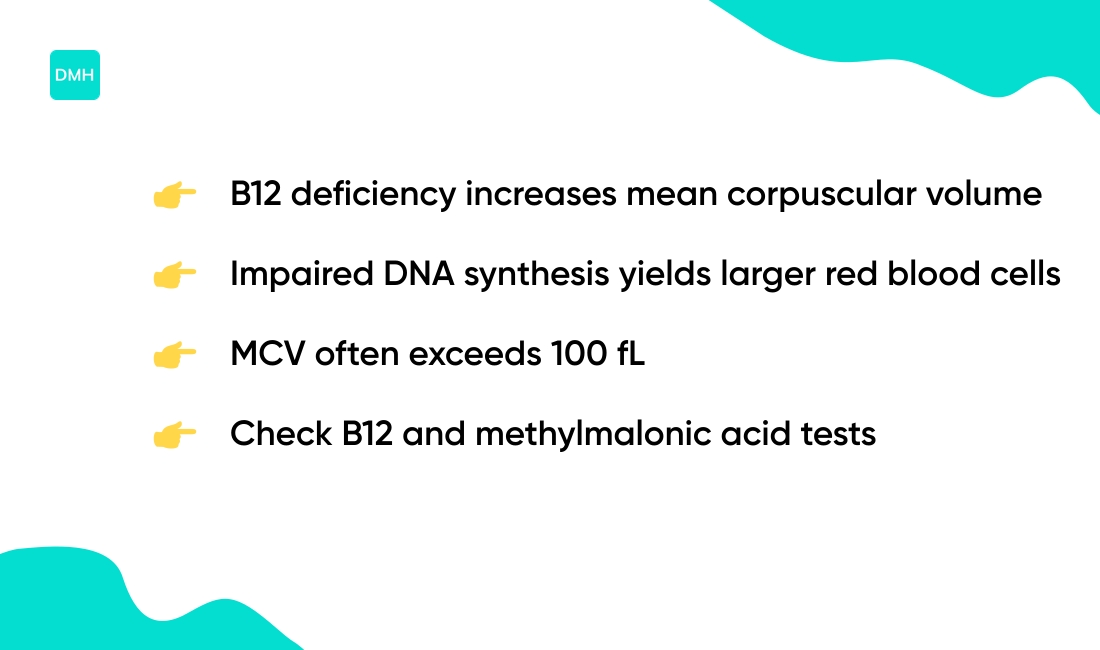
B12 deficiency raises mean corpuscular volume by impairing DNA synthesis in red blood cell precursors. Delayed DNA replication forces cells to grow without dividing.
That process produces larger red cells and macrocytosis on an MCV blood test. MCV in CBC often climbs above 100 fL with true deficiency.
Mild cases sit near 100–110 fL. Severe or long-standing deficiency and pernicious anemia can push MCV past 110 fL. Pernicious anemia causes autoimmune B12 loss and shows a clear link to marked macrocytosis.
Checking methylmalonic acid and B12 levels gives the best confirmation. If you see high MCV values on a CBC, ask your clinician about B12 testing and follow-up labs to guide treatment.
Folate deficiency and MCV levels
Folate supports DNA synthesis in bone marrow. Without it, precursor cells fail to divide—cells enlarge instead of multiplying. That change causes macrocytic anemia and raises mean corpuscular volume.
Dietary sources include leafy greens, legumes, fortified cereals, and liver. Adults need about 400 mcg folate daily.
On a CBC, MCV can rise before hemoglobin drops. MCV often exceeds 100 fL with ongoing deficiency, and severe lack can push MCV above 110 fL. Reticulocyte response usually stays low during early recovery.
Routine MCV review catches many nutrient gaps. You should order folate and B12 tests if MCV is high, then adjust diet or start supplementation as guided by results.
Other causes of elevated MCV
Elevated mean corpuscular volume can come from many sources beyond B12 or folate deficiency:
- Chronic alcohol use damages marrow and inflates red cell size
- Liver disease effects alter lipid membranes and raise MCV
- Hypothyroidism slows red cell turnover and lifts MCV slightly
- Certain medications—like anticonvulsants and zidovudine—can raise MCV
- Myelodysplastic syndromes and other marrow disorders cause marked increases
- Aging often nudges MCV upward after age 25
Clinicians should review meds, liver tests, and thyroid function. Don’t assume nutritional causes first. (I’ve seen alcohol-related MCV rises of 6–12 fL in practice.)
You can read detailed data at OptimalDx MCV research.
How to interpret MCV on a CBC
We treat mean corpuscular volume as a core clue for anemia classification. MCV reports average red blood cell size in femtoliters (fL).
Labs usually use a range of about 80–100 fL. Values below 80 fL or above 100 fL require context.
Low MCV (<80 fL) indicates microcytic anemia. Common causes include iron deficiency and thalassemia. If MCH also falls, iron deficiency is more likely; if RDW stays normal, thalassemia trait becomes more likely.
High MCV (>100 fL) points to macrocytic anemia. B12 or folate deficiency often explains this pattern. Chronic alcohol use, liver disease, and some drugs also raise MCV.
| MCV Range | Classification | Common Causes | Confirmatory Tests |
|---|---|---|---|
| <80 fL | Microcytic | Iron deficiency, thalassemia | Ferritin, TIBC, hemoglobin electrophoresis |
| 80–100 fL | Normocytic | Chronic disease, kidney disease, acute blood loss | Reticulocyte count, renal panel, hemolysis markers |
| >100 fL | Macrocytic | B12/folate deficiency, alcohol, liver disease | B12, folate, methylmalonic acid, liver function tests |
Use MCH to see hemoglobin per cell in picograms. Use MCHC to judge hemoglobin concentration in each cell. An RDW over about 15% shows wide size variation and suggests mixed causes or recent recovery.
Here’s a pattern example: MCV 72 fL, MCH 23 pg, RDW 18% fits iron deficiency. MCV 105 fL with low reticulocytes fits B12 or folate lack.
Check reticulocyte response via reticulocyte count. A brisk reticulocyte rise means blood loss or hemolysis; a poor reticulocyte response means production failure.
Reading MCV with RDW, MCH, and reticulocytes gives faster answers than any single number. You should ask your clinician for targeted tests like ferritin, B12, and folate, then track trends over weeks.
What is the difference between MCV and MCH
MCV and MCH measure different red blood cell features. MCV shows cell size; MCH shows hemoglobin content per cell.
The mean corpuscular volume records average red blood cell size. Labs list it in femtoliters (fL). Typical adult range sits near 80–100 fL.
The mean corpuscular hemoglobin gives the hemoglobin mass inside each cell. Labs report it in picograms (pg). Normal MCH usually falls between 27 and 33 pg.
When values diverge, interpretation matters. Low MCV with low MCH points to microcytic, hypochromic anemia—iron deficiency or thalassemia often cause that pattern.
Normal MCV with low MCH suggests small hemoglobin stores per cell. High MCV with normal or low MCH suggests macrocytic cells with reduced hemoglobin concentration.
Use MCV and MCH with RDW and MCHC for a clear picture. If you see MCV 76 fL and MCH 25 pg, suspect iron deficiency and check ferritin. If MCV is 105 fL with low MCH, check B12 and folate and consider a smear.
Ask your clinician for ferritin, B12, folate, and a peripheral smear when MCV and MCH conflict. Reading both values together stops simple misreads—you’ll get better answers when you bring these numbers to your appointment.
What does normal MCV with anemia mean
Mean corpuscular volume measures average red blood cell size. A normal MCV with low hemoglobin defines normocytic anemia. Your red cells look normal in size but carry too little hemoglobin or exist in low numbers.
Common normocytic anemia causes include:
- Chronic inflammatory disease—inflammation alters iron handling and shortens red cell lifespan
- Kidney disease with low erythropoietin—lowers red cell production
- Acute blood loss—counts drop rapidly while cell size stays normal
- Hemolysis or marrow failure—production and survival both suffer
Follow-up testing matters. Order a reticulocyte count, iron studies, renal panel, and markers of hemolysis. Consider bone marrow evaluation when tests remain unclear.
Check the page on what type of cancer causes low hemoglobin if you suspect malignancy as a cause of anemia.
Prompt evaluation is important when hemoglobin is under 10 g/dL or when you feel dizzy, tired, or short of breath. Treat the underlying condition rather than focusing only on numbers. If you need help interpreting results, bring them to your clinician and ask for these specific tests.
Can dehydration change MCV results
Yes, dehydration can change mean corpuscular volume results by concentrating blood. We recommend hydrating before a CBC to lower the risk of false elevation.
Low fluid intake reduces plasma. That raises red cell proportion in the sample. Lab instruments calculate MCV from hematocrit and red blood cell count using MCV = (Hct ÷ RBC) × 10 fL. Plasma shifts can alter MCV by a few femtoliters.
Small shifts matter when your result lies near a cutoff. If MCV sits near the lab’s upper limit, dehydration can push it above the threshold for macrocytosis.
Here’s a practical step: drink 500 mL water about 15 to 30 minutes before the draw unless your clinician advises otherwise. That simple action often reduces hemoconcentration and gives a truer reading.
If you suspect dehydration, repeat testing after rehydration. A repeat sample can distinguish true high MCV from a dilution effect due to plasma volume loss.
Keep a record of symptoms like dizzy lightheadedness or reduced urine. Share these with your provider when discussing an abnormal MCV—accurate interpretation helps guide the right next step for you. Ask your clinician to repeat the test if uncertain.
You’ll also like: What Is Neutrophils In Blood Test
Health implications of abnormal MCV levels
Shifts in MCV are more than a lab quirk. The mean corpuscular volume measures average red blood cell size in femtoliters. Normal MCV range runs near 80 to 100 fL, though labs may use slightly different cutoffs.
Recent research links MCV to broader health risks:
- MCV above 95 fL links to faster cognitive decline in cohort studies
- Higher MCV associates with arterial stiffening and reduced vascular elasticity
- Elevated MCV connects to insulin resistance and metabolic strain
- Patients with chronic disease and high MCV show higher mortality rates
- Stroke outcomes show a U-shaped relation with MCV, with risks at low and high extremes
If your MCV rises past 95 fL, ask your clinician about a referral to a hematologist for targeted evaluation.
Mild MCV shifts deserve prompt follow-up. (I’ve seen early intervention clarify nutrient gaps and medication effects.) Start with a repeat CBC, iron studies, B12, and folate levels within four to eight weeks. Adjust treatment to the underlying cause and monitor MCV levels over time.
You might also like: Can Low Iron Cause Headaches
How to treat abnormal MCV levels
Targeted care speeds recovery and limits unnecessary tests.
For low MCV, often from iron deficiency, start iron replacement. Oral ferrous sulfate 325 mg supplies about 65 mg elemental iron. Many clinicians prefer 65 mg elemental iron daily or every other day to reduce side effects.
Expect a reticulocyte rise within 7–10 days and hemoglobin to increase about 1 g/dL every 2–4 weeks.
For high MCV from B12 or folate lack, give replacement. B12 can work as 1,000 mcg intramuscular weekly until levels normalize, then monthly. High-dose oral B12 (1,000–2,000 mcg daily) helps some patients. Folate at 1 mg daily corrects dietary deficiency fast. Watch for neurologic signs with low B12.
Address chronic conditions that raise MCV. Treat alcohol use, liver disease, hypothyroidism, and review medicines that affect red cell size. For low hematocrit or persistent anemia, consider steps to raise red cell mass; see how to raise hematocrit for practical options.
When should you seek urgent care? Seek evaluation when symptoms are severe, when hemoglobin falls below 8 g/dL, or when neurologic changes appear. Prompt specialist review gives the best chance to correct the issue.
Read also: Does Anemia Cause Headaches
Educational notice: This content is provided for informational and educational purposes only and is not intended as medical advice. Always consult a qualified healthcare professional for medical concerns.