
A single glance at two different colored eyes often sparks curiosity.
Heterochromia describes a difference in iris pigmentation between the two eyes or within one iris.
People frequently wonder whether it’s just a cosmetic quirk, a hereditary trait, or something that needs medical attention.
Daily Medical Health breaks down causes (genetic and acquired), types like complete, sectoral and central heterochromia, related syndromes, and when to seek evaluation.
You’ll learn how eye color genetics and melanin distribution create different colored eyes, what signs suggest underlying disease, and the diagnostic and cosmetic options available.
What is heterochromia?
Heterochromia iridum is defined as a variation in iris color between the two eyes or within a single iris. The iris is the colored ring that controls how much light enters the eye.
Eye color depends on melanin, a brown pigment produced by cells in the iris. Higher melanin levels produce brown tones, while lower melanin levels produce blue or green tones.
Uneven melanin pigment distribution can create different colored eyes or distinct color segments inside one eye. Genetic factors influence melanin production and can cause congenital heterochromia.
Genes such as OCA2 and HERC2 may play a role in eye color genetics. Actually, research suggests at least 50 genes contribute to iris hue, making inheritance patterns quite complex.
In some people, changes to melanin after injury, inflammation, or medication can lead to acquired heterochromia. Presentation ranges from a full-color difference between eyes to a small colored sector or a central ring.
Many cases don’t affect vision and remain a cosmetic feature. Some cases may signal an underlying condition and warrant evaluation.
Always consult a qualified healthcare professional for medical advice specific to your situation. Not medical advice; content for educational purposes, consult a professional.
Types of heterochromia
Complete heterochromia
Complete heterochromia is a form of heterochromia iridum where one iris shows a different color from the other. You may see one blue eye and one brown eye.
It’s usually benign and congenital. Melanin distribution in each iris determines color, with genetic factors influencing melanin during fetal development.
Estimates place occurrence under 1% of people, though figures vary across studies. Vision typically remains normal with this condition.
Some cases arise after injury or disease and may indicate an underlying issue. If your eye color changes later in life, an eye evaluation may identify causes.
This trait appears in some animals more often than in people. The pattern may be hereditary or result from mosaicism.
Always consult a qualified healthcare professional for medical advice specific to your situation. Not medical advice; content for educational purposes, consult a professional.
Sectoral heterochromia
Sectoral heterochromia is a type of heterochromia iridum where a sector or wedge of one iris shows a different color from the rest. This form is often called partial heterochromia.
Iris color reflects local melanin levels. A localized change in melanin produces the color difference, creating a striking, patchy look within one eye.
It may be present at birth due to genetic variation. Plus, it can appear after trauma, inflammation, or medication.
This sectoral heterochromia iris pattern often creates a multicolored single eye effect. Congenital forms generally don’t affect vision.
New-onset sectoral heterochromia may prompt an eye exam to check for underlying causes. Always consult a qualified healthcare professional for medical advice specific to your situation.
Not medical advice; content for educational purposes, consult a professional.
Central heterochromia
Central heterochromia is defined as a ring of different pigment around the pupil that contrasts with the outer iris. The inner ring often appears lighter or darker than the surrounding iris.
Melanin concentration varies across the iris and can create a sharp color break near the pupil. A distinct inner ring marks central heterochromia, while the outer iris boundary often stays a single color.
This helps distinguish it from hazel eyes where tones blend without a clear ring. Have you noticed a defined color boundary in your own eyes?
Central heterochromia may be congenital or develop later from changes in pigment. Frequency varies across populations and between species such as cats and dogs.
Research on exact causes links melanin distribution and genetics, though findings can vary. More detail appears at HowStuffWorks.
Always consult a qualified healthcare professional for medical advice specific to your situation. Not medical advice; content for educational purposes, consult a professional.
What causes heterochromia?
Genetic and congenital causes
Heterochromia is a difference in iris color between eyes or within one iris. Genes such as OCA2 and HERC2 regulate melanin production in the iris.
Variants in these genes can lower or raise pigment levels and alter color. Some benign mutations present at birth may cause one eye to be blue and the other brown.
Such cases often pose no vision risk. Eye color follows a complex, polygenic pattern, with genetic studies estimating at least 50 genes influencing iris hue.
Interactions between many loci can produce partial, central, or complete heterochromia. Interestingly, 10-20% of children experience shade changes up to age 6, and 15% of white adults see hue shifts over time.
Genetic testing can clarify inheritance in some families, though results vary and don’t always predict exact color. If you notice a new change in eye color, seek evaluation.
Always consult a qualified healthcare professional for medical advice specific to your situation. Not medical advice; content for educational purposes, consult a professional.
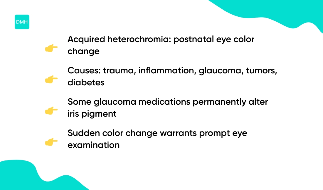
Acquired heterochromia from injury or disease
Acquired heterochromia is a change in eye color that develops after birth. You may notice one eye darkening or a colored sector appearing in one iris.
Common causes include:
- Eye trauma or surgery
- Inflammation such as uveitis or iritis
- Glaucoma and its medications
- Tumors (benign or cancerous)
- Diabetes complications
- Fuchs’ heterochromic cyclitis
- Pigment dispersion syndrome
Some glaucoma drugs alter pigment and may produce long-term changes. These changes can signal underlying disease and warrant an eye exam.
Learn more about signs of iris inflammation by reading iritis vs conjunctivitis for comparison. Findings vary by cause and by person; diagnostic tests often include slit-lamp exam and imaging.
Well, a recent color change deserves prompt evaluation even if vision remains normal. Always consult a qualified healthcare professional for medical advice specific to your situation.
Not medical advice; content for educational purposes, consult a professional.
Medication-induced heterochromia
Certain medicines can change iris color over time. Eye drops called prostaglandin analogues have the clearest link (commonly prescribed for glaucoma management).
These drops can stimulate iris melanocytes, causing an iris pigment increase and gradual darkening. Color change may move blue or hazel eyes closer to brown.
Reports vary, but generally under 10% of patients notice visible change after months to years. The effect often appears only in the treated eye, producing acquired heterochromia iridum.
Some patients see permanent change. FDA labeling for latanoprost and similar drugs lists iris pigmentation as a reported adverse effect.
An eye care specialist can explain cosmetic options and monitoring. Always consult a qualified healthcare professional for medical advice specific to your situation.
Not medical advice; content for educational purposes, consult a professional.
Syndromes and conditions linked to heterochromia
Heterochromia iridum refers to different colored eyes or distinct color zones within one iris. It can occur alone or with broader pigmentary differences linked to genetic syndromes and neurological conditions.
Waardenburg syndrome often shows patchy pigment changes. You may see total or sectoral heterochromia alongside hair and skin pigment differences.
Horner’s syndrome can produce lighter iris color on the affected side when it develops early in life. The change reflects sympathetic nerve disruption that alters pigment over time.
Other conditions associated with heterochromia include:
- Sturge-Weber syndrome with vascular growths
- Tuberous sclerosis with hamartomatous changes
- Piebaldism causing white skin patches and sectoral heterochromia
- Neurofibromatosis with iris Lisch nodules
- Hirschsprung disease in rare presentations
- Bloch-Sulzberger syndrome
Acquired disorders such as ocular inflammation, tumors, or medication effects can create new heterochromia later in life. Some rare metabolic and chromosomal conditions also list heterochromia among findings.
| Condition | Type of Heterochromia | Associated Features |
|---|---|---|
| Waardenburg syndrome | Complete or sectoral | Hair/skin pigment changes, hearing loss |
| Horner’s syndrome | Lighter affected iris | Ptosis, miosis, anhidrosis |
| Sturge-Weber | Sectoral | Facial port-wine stain, glaucoma |
| Piebaldism | Sectoral | White forelock, skin depigmentation |
Clinical evaluation often includes eye exam, medical history, genetic testing, and imaging when a syndrome is suspected. A peer-reviewed overview links pigmentary signs with systemic conditions at Oxford Academic.
Findings vary by patient and by condition. Always consult a qualified healthcare professional for medical advice specific to your situation. Not medical advice; content for educational purposes, consult a professional.
How rare is heterochromia?
Heterochromia is uncommon in people. Estimates place heterochromia in humans under 1% of the global population, with complete heterochromia appearing even rarer.
Sectoral and central heterochromia occur more often than complete forms. Prevalence can vary by ancestry and genetic background.
Some clinical series suggest higher rates in groups with pigment-related variants. Heterochromia genetics involve multiple genes affecting melanin production and distribution.
Heterochromia in animals appears more common than in humans. Cats with white coat genes often show different colored eyes, while certain dog breeds display notably higher rates.
Here’s the thing: Siberian Huskies, Australian Shepherds, and Dalmatians show increased heterochromia frequency. Breed studies estimate rates above 10% in selected dog groups.
Horses with pinto or splashed white patterns may display sectoral heterochromia. Animal heterochromia links to coat-color genes such as piebald and merle.
Human heterochromia can be congenital or acquired from injury or disease. Did you know acquired color change may signal underlying eye disease?
Evidence varies and study methods differ across reports, so findings can be inconsistent. Always consult a qualified healthcare professional for medical advice specific to your situation.
Not medical advice; content for educational purposes, consult a professional.
Is heterochromia dangerous?
Congenital heterochromia is often benign and usually doesn’t affect vision. Color differences reflect melanin distribution in the iris, with melanin levels varying by genetics.
Acquired heterochromia that appears later in life may be a sign of another condition. Possible causes include eye inflammation, injury, pigmentary glaucoma, tumors, and some glaucoma medications.
A new brown spot on the iris can matter. See brown spot on eye for related information.
An eye specialist will review medical history and examine the eye through slit-lamp exams. They may order blood tests or imaging to rule out systemic issues.
Findings can vary by case. Management targets the underlying cause when one exists, while cosmetic options such as colored contacts may address appearance alone.
For most people with congenital heterochromia, risk remains low. But new or changing color deserves evaluation (better safe than sorry).
Always consult a qualified healthcare professional for medical advice specific to your situation. Not medical advice; content for educational purposes, consult a professional.
Read also: How To Tighten Eyelid Skin Without Surgery
How is heterochromia diagnosed?
Diagnosis of heterochromia aims to find the cause of different colored eyes. Clinicians begin with a comprehensive eye examination and record a patient medical history noting onset, trauma, medications, and systemic symptoms.
Exam steps include:
- Slit-lamp inspection of the iris
- Pupil testing for symmetry and reaction
- Anterior segment photography
- Intraocular pressure measurement
- Gonioscopy to inspect drainage angle
- Posterior segment exam via dilated fundoscopy
- Optical coherence tomography for retina and optic nerve
Intraocular pressure readings above 21 mm Hg may suggest glaucoma. Gonioscopy inspects the drainage angle for pigment or abnormal structures.
If a mass or deep lesion is suspected, ultrasound B-scan or cross-sectional imaging is ordered. MRI or CT can help identify orbital tumors or vascular malformations that change iris color.
Blood tests may screen for infectious or inflammatory causes such as syphilis or autoimmune markers. Some acquired cases trace to prostaglandin eye drops or uveitis; congenital cases often show no pathology.
For instance, a person with one blue and one brown eye who has no other signs of disease typically needs no further workup. Evidence varies by presentation; workup remains individualized and guided by clinical findings.
Always consult a qualified healthcare professional for medical advice specific to your situation. Not medical advice; content for educational purposes, consult a professional.
Read also: How to Remove Dark Circles Under Eyes Permanently
Treatment options for heterochromia
Congenital heterochromia usually needs no medical treatment since vision often remains normal. Acquired heterochromia can indicate injury, inflammation, medication effects, or systemic disease.
Evaluation aims to find the underlying cause through eye exam, slit-lamp inspection, blood tests, or imaging. Management depends on the diagnosis.
Treatment approaches vary based on cause:
- Inflammatory conditions may respond to anti-inflammatory therapy
- Tumors or vascular problems may require specialist care
- Medication-induced color change may stabilize after stopping the drug
- Some pigment changes can persist even after treatment
Colored contact lenses offer a noninvasive cosmetic option when appearance matters. Lenses come in prescription and plano forms, with proper fitting and hygiene reducing infection risk.
For context, prostaglandin eye drops used for glaucoma can darken the iris over months in some people. That change may not reverse after stopping the drops.
Outcomes vary by cause, duration, and individual factors. Some acquired changes resolve with treatment, while other changes remain permanent.
If you notice a sudden color change or new visual symptoms, seek professional evaluation promptly. Always consult a qualified healthcare professional for medical advice specific to your situation.
Not medical advice; content for educational purposes, consult a professional.
You’ll also like: What Causes Sudden Temporary Double Vision?
Educational notice: This content is provided for informational and educational purposes only and is not intended as medical advice. Always consult a qualified healthcare professional for medical concerns.