
Noticing your ultrasound says “anterior placenta” can feel worrying when you don’t know what it means.
Many expectant parents worry about delayed fetal movement, monitoring changes, or delivery risks.
(And that’s completely understandable!)
This article explains what an anterior placenta is, why it happens, and how it may change what you feel and how providers monitor the baby.
You’ll learn when kicks are usually felt with this placenta position, how it shows up on ultrasound, and when to contact your clinician.
It also covers potential complications like low-lying placenta or placenta previa, how the placenta can shift as the uterus grows, and delivery considerations.
By the end you’ll be better prepared to track movement, understand scan results, and discuss delivery options confidently with your care team.
What is an anterior placenta?
An anterior placenta forms when the placenta implants on the front uterine wall.
The placenta connects the developing fetus to the uterine lining and provides oxygen and nutrients throughout pregnancy.
Placenta position varies widely. Common placements include anterior, posterior, fundal, and lateral.
An anterior placement sits between the fetus and the abdominal wall. This position can cushion fetal movement and alter how kicks feel through your belly.
Some reports estimate an anterior placenta occurs in about 20% of pregnancies, though prevalence can vary by study and population. The finding is a normal anatomical variation in most cases.
Have you wondered how doctors identify placenta location? Ultrasound imaging identifies placenta position early in prenatal care. Technicians note anterior placement on routine scans and track its position as the uterus grows.
An anterior placenta doesn’t inherently indicate a problem. Most people with this placement have healthy pregnancies and deliveries. Certain specific concerns, such as a low-lying anterior placenta or placenta previa, require monitoring.
Clear communication with your prenatal clinician helps interpret scan results and follow-up plans. If you’re concerned about placenta position, ask your care team for clarification and monitoring steps.
Not medical advice; content for educational purposes only. Always consult a qualified healthcare professional for medical advice specific to your situation.
What causes an anterior placenta?
An anterior placenta forms when the placenta implants on the front wall of the uterus.
Placenta position reflects where the fertilized egg attaches to the uterine lining at conception. This implantation process occurs largely by chance and lies outside voluntary control.
Some studies report anterior placentas in about 20–30% of pregnancies, suggesting it’s a common variation.
Certain uterine features may be associated with where implantation happens. Scarring from prior surgery, uterine fibroids, and natural variations in uterine shape can influence the implantation site.
Biological processes guide early embryo attachment. Local blood flow, endometrial receptivity, and microscopic tissue signals play a role.
Here’s the thing: placental location can shift as the uterus grows. Low-lying placentas often migrate upward during the second trimester.
An anterior placenta doesn’t determine fetal sex and doesn’t imply specific outcomes by itself. Most people with this placement have routine pregnancies and births.
If you have questions about placenta placement, mention them at a prenatal visit so your clinician can review ultrasound findings.
Not medical advice; content for educational purposes only. Always consult a qualified healthcare professional for medical advice specific to your situation.
How does an anterior placenta affect feeling baby kicks?
When will I feel fetal movement with an anterior placenta?
An anterior placenta implants on the front wall of the uterus and can cushion fetal movement, which may delay when kicks are felt.
Most people notice fetal movement after 20 weeks. Sensation commonly appears between 20 and 24 weeks with an anterior placement.
Some people feel movement as early as 16 weeks, especially after prior pregnancies. But with an anterior placenta, you might not feel those early flutters quite as soon.
The placenta acts as a soft buffer. Movements may feel gentler against the belly with this position.
Wondering why your doctor has trouble finding the heartbeat? Doppler heartbeat checks can be less sensitive early with an anterior placenta, as the placental tissue dampens the sound.
If movement seems reduced after 28 weeks, clinical assessment may be appropriate. Tracking patterns helps you notice changes that warrant discussion with your care team.
Not medical advice; content for educational purposes only. Always consult a qualified healthcare professional for medical advice specific to your situation.
Does an anterior placenta affect kick counts and monitoring?
An anterior placenta sits on the front uterine wall and acts as a soft layer between you and the baby. The placental tissue can mask movements and make kicks feel gentler.
Feeling baby move may occur later with an anterior placenta. Most people notice movement by 18–20 weeks. With anterior placement, detection often shifts to about 20–24 weeks.
Kick counting can feel less clear. Maternal perception may undercount smaller, inner movements. Some guidance suggests aiming for ten movements within two hours during focused checks.
Well, marked drops in movement may signal a need for clinical assessment. Monitoring plans can vary from person to person and often include routine clinic checks or fetal testing if concerns arise.
Not medical advice; content for educational purposes only. Always consult a qualified healthcare professional for medical advice specific to your situation.
What does an anterior placenta look like on ultrasound?
The placenta sits along the anterior uterine wall and appears as a smooth, echogenic band between the fetus and the abdominal wall.
Technicians view it on transabdominal scans. They use transvaginal ultrasound when the placental edge needs clearer detail.
Color Doppler highlights placental vessels and confirms location. Sonographers measure the placental edge from the internal os in centimeters.
A low-lying anterior placenta sits near or over the cervix. Some guidelines note a placental edge within 2 cm of the internal os as low-lying.
Placental thickness at the anterior wall may range about 2 to 4 cm, depending on gestational age. Reports typically state “anterior placenta” and give the edge distance in centimeters.
Placental appearance varies with maternal anatomy and fetal position. Not medical advice; content for educational purposes only. Always consult a qualified healthcare professional for medical advice specific to your situation.
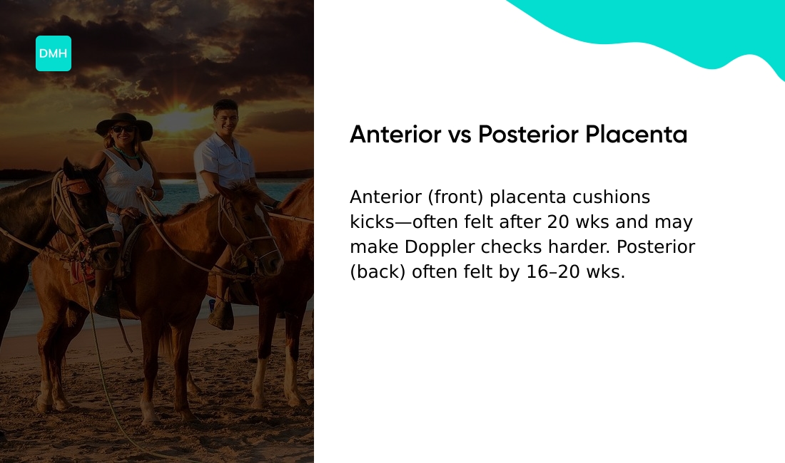
Anterior placenta vs posterior placenta
The anterior placenta position means the placenta implants on the front wall of the uterus. The posterior placenta position means it attaches to the back wall.
Both placements are common and usually normal.
Estimates suggest about 1 in 5 pregnancies have an anterior placenta. Placenta location sets early, at implantation, and it often changes as the uterus grows.
The main difference lies in sensation. An anterior placenta may act as a cushion, so you may detect fetal movement later, often after 20 weeks. A posterior placenta often lets movements feel sooner, sometimes by 16 to 20 weeks.
Curious how this affects daily monitoring? This affects feeling baby movement and kick counts throughout your pregnancy.
Ultrasound shows placenta location clearly. Technicians note front or back placement during routine scans. An anterior placenta can make early heartbeat checks with a Doppler harder to hear.
|
Feature |
Anterior Placenta |
Posterior Placenta |
|---|---|---|
|
Location |
Front uterine wall |
Back uterine wall |
|
Movement Detection |
Often after 20 weeks |
Often 16–20 weeks |
|
Doppler Detection |
May be harder early |
Typically easier |
|
Health Concerns |
Usually normal; monitor if low-lying |
Usually normal |
Most research finds no consistent health advantage for either position. Low-lying anterior placentas may raise placenta previa concerns and need follow-up.
Reliable patient information appears on the Cleveland Clinic site about anterior placenta.
Both positions can lead to healthy outcomes. If you’re concerned about movement changes or bleeding, consider speaking with your clinician.
Not medical advice; content for educational purposes only. Always consult a qualified healthcare professional for medical advice specific to your situation.
Does an anterior placenta increase pregnancy risks?
Can an anterior placenta be low-lying?
A placenta that sits on the front wall can also lie low near the cervix. This is called a low-lying anterior placenta.
It differs from a typical anterior placement that rests higher on the uterine front wall.
Uterine growth can shift the placenta upward as pregnancy progresses. Some studies report that up to 80–90% of low placentas seen on early scans move away from the cervix by late pregnancy, according to available research.
This process is often called placental migration. A low front placenta can raise the placenta previa risk if it covers the cervical opening.
Ultrasound helps track the anterior placenta position over time. If bleeding or new symptoms occur, clinical follow-up is commonly advised.
Not medical advice; content for educational purposes only. Always consult a qualified healthcare professional for medical advice specific to your situation.
What is anterior placenta previa?
Anterior placenta previa occurs when the placenta implants on the front uterine wall and partially or fully lies over the cervical opening.
When the placenta covers the cervix, it can block the birth canal and may cause bleeding in later pregnancy.
People may notice painless vaginal bleeding. Bleeding ranges from light spotting to heavier loss.
Clinicians identify and monitor this condition with obstetric ultrasound. Repeat scans occur in the second and third trimester to check whether the placenta moves as the uterus grows.
Some studies suggest placenta previa affects about 0.3–0.5% of pregnancies at term. Risk appears higher after prior cesarean or with multiple pregnancies.
Painless vaginal bleeding often prompts urgent evaluation. If you experience this symptom, contact your healthcare provider promptly.
Not medical advice; content for educational purposes only. Always consult a qualified healthcare professional for medical advice specific to your situation.
Can the placenta move during pregnancy?
The placenta can appear to move as the uterus grows. Uterine expansion stretches the wall and makes the attachment look higher.
A low-lying anterior placenta may rise away from the cervix without intervention. Some studies report more than 80% of low-lying placentas found at 20 weeks resolve by 34 weeks.
Clinicians track placenta position changes with ultrasound. Routine anatomy scans at 18 to 22 weeks record location and note any low placement.
Clinicians schedule repeat scans in the third trimester if the placenta sits near the cervix. If the placenta still covers the cervix late in pregnancy, providers discuss delivery options.
Anterior placenta migration describes this apparent shift during growth. Position can vary from person to person and may be monitored until delivery.
Not medical advice; content for educational purposes only. Always consult a qualified healthcare professional for medical advice specific to your situation.
Anterior placenta labor and delivery
Most pregnancies with an anterior placenta proceed to a normal vaginal birth. The placenta sits on the front uterine wall and rarely blocks the birth canal.
An anterior placenta can cushion labor sensations. Early labor pulses and first contractions may feel different, though external monitoring remains effective in most cases.
Fetal position affects labor more than placenta position. An anterior placenta may be linked to a higher chance of a back-to-back baby or occiput posterior position, according to some observational studies.
That position can lengthen labor and raise the likelihood of assisted delivery or conversion to a cesarean delivery.
Low-lying anterior placenta or placenta previa changes delivery planning. Placenta previa occurs in under 1% of pregnancies at term. If the placenta covers the cervix, most clinicians plan a cesarean delivery to avoid major bleeding.
Placenta accreta spectrum is uncommon but more likely when the placenta overlies prior uterine scars. Imaging and repeat scans guide management if concern exists.
Labor monitoring typically includes continuous or intermittent fetal heart assessment. Care teams consider bleeding, fetal status, and labor progress when deciding mode of delivery.
If you’re concerned about positioning or delivery options, discuss imaging results and birth plans with your clinician. Evidence varies and outcomes can vary from person to person.
Not medical advice; content for educational purposes only. Always consult a qualified healthcare professional for medical advice specific to your situation.
Do I need a C-section with an anterior placenta?
Most people with an anterior placenta can plan for a vaginal birth. The placenta sits on the front uterine wall, and that placement alone rarely forces a cesarean.
Cesarean delivery may be needed if the placenta covers the cervix. This condition, called placenta previa, can cause bleeding and often leads to planned C-section.
A low-lying anterior placenta that remains near the cervix into the third trimester may increase the chance of cesarean.
Other factors affect delivery method. A prior uterine surgery, persistent bleeding, fetal distress, or a malpositioned baby can lead to cesarean.
Providers use ultrasound to check placenta location and fetal position during the second and third trimesters. An anterior placenta ultrasound often shows a clear front-wall placenta image.
Feeling baby moves may differ with an anterior placenta. The placenta can cushion fetal movement, which may delay kick awareness. That difference doesn’t predict delivery type.
Kick counts and fetal monitoring remain useful for tracking fetal well-being.
Discussing findings with your clinician helps clarify options. Decisions vary from person to person and consider placenta position, bleeding, prior surgeries, and labor progress.
If you’re concerned, consider speaking with a clinician who can review your scans and history.
Not medical advice; content for educational purposes only. Always consult a qualified healthcare professional for medical advice specific to your situation.
You might also like: How To Get Doctor To Extend Maternity Leave
Common myths about anterior placenta
The placenta that attaches to the front wall of the uterus may spark many myths. People often link placement to fetal traits and pregnancy outcomes.
One common anterior placenta myth claims that placenta position predicts baby sex. Research finds no reliable link between placenta location and gender.
Myths about anterior placenta kicks say a soft or late-feeling bump means a weak baby. Actually, the placenta can cushion movements, which often delays feeling kicks until after 20 weeks. Delayed fetal movement detection doesn’t indicate a problem by itself.
Anterior placenta ultrasound myths suggest ultrasound images reveal personality or future health. Ultrasound shows location and health markers. Clinicians use scans to monitor growth and position, not personality traits.
Some sources claim anterior placentas raise common pregnancy complications. Some studies suggest small associations with conditions like hypertension or gestational diabetes, but findings vary and may reflect other factors.
Have you heard claims about belly shape predicting outcomes? Stories on forums can spread anecdotal links between belly shape, sleeping position, and outcomes. Scientific evidence doesn’t support consistent predictions from belly shape or posture.
Emotional stress can follow belief in these myths. If relationship strain or emotional worry occurs, reading about feeling rejected by husband during pregnancy may offer perspective and coping ideas.
Here’s the bottom line: anterior placenta often represents a normal variation. Patterns vary from person to person and require clinical context.
Not medical advice; content for educational purposes only. Always consult a qualified healthcare professional for medical advice specific to your situation.
Read also: I Accidentally Gave My Newborn Water
When to contact your doctor
Urgent signs in pregnancy may need prompt medical evaluation.
-
Vaginal bleeding. Any new bleeding that’s heavy or persistent may be serious. Bleeding that soaks one pad per hour for several hours is commonly treated as urgent by clinicians.
-
Reduced fetal movement. A noticeable drop in baby movement after 20 weeks can be concerning. If movements fall over a 12-hour period, consider contacting a clinician for assessment.
-
Severe abdominal or back pain. New, intense pain that doesn’t ease may indicate a complication and may require evaluation.
-
Constant contractions. Regular, painful contractions before 37 weeks may suggest preterm labor. Contractions every 10 minutes or closer for an hour often prompt clinical review.
-
Fever or fluid leakage. A fever above 100.4°F (38°C) or clear fluid leaking from the vagina can signal infection or rupture of membranes.
An anterior placenta can cushion fetal movement and may delay when you feel kicks, often until after 20 weeks. Cushioning doesn’t remove the need to monitor activity.
If you have concerns about movement patterns, discuss them with your care team. Read more about unusual belly sensations at water sound in belly during pregnancy.
Regular prenatal check-ups help monitor placenta position, fetal growth, and maternal health. If you’re concerned about any symptom, consider speaking with a clinician for timely evaluation.
Not medical advice; content for educational purposes only. Always consult a qualified healthcare professional for medical advice specific to your situation.
You might also like: Prenatal Vitamin Mechanism Of Action
Educational notice: This content is provided for informational and educational purposes only and is not intended as medical advice. Always consult a qualified healthcare professional for medical concerns.