
A quick, noninvasive scan can reveal heart issues that a physical exam or EKG might miss.
What is an echocardiogram and why might you need one?
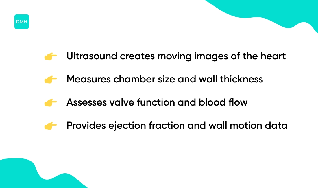
Well, it’s a cardiac ultrasound (echo heart test) that uses sound waves to create moving images of heart chambers, valves, pumping strength and blood flow.
People often face vague symptoms—shortness of breath, chest pain or palpitations—and need clearer answers than other tests provide.
In this guide you’ll learn the main echocardiogram types (transthoracic, transesophageal, stress, Doppler, 2D/3D), how the procedure’s performed, and what results can show.
By the end you’ll understand what to expect from the echocardiogram test and how it helps doctors guide treatment.
What is an echocardiogram
An echocardiogram is a noninvasive heart test that uses ultrasound. High-frequency sound waves travel from a handheld transducer into the chest.
Echoes reflect off the heart and return to the transducer. The device converts echoes into real-time images on a monitor.
Physicians observe valves, chambers, and blood flow. Clinicians often call this exam an echo heart test or cardiac ultrasound test.
It shows heart size, pumping strength, valve motion, and major structural abnormalities. The test reports ejection fraction, a percentage that estimates pumping efficiency.
The procedure typically takes 15 to 60 minutes. Technicians apply gel and move the transducer across the chest wall. The exam causes no radiation and usually causes no pain.
Echocardiography can include Doppler analysis to measure blood flow speed and direction. Specialized methods provide two-dimensional or three-dimensional views.
Clinicians use the test to evaluate chest pain, shortness of breath, or abnormal heart sounds. Pediatric echocardiogram adapts the technique for infants and children. Image quality depends on body habitus and operator skill.
Abnormal results may prompt further testing such as cardiac catheterization or CT angiography. Evidence indicates echocardiograms detect structural problems well but can vary in detecting coronary blockages.
Not a medical advice, content for educational purposes, consult a professional.
How does an echocardiogram work
An echocardiogram uses sound to image the heart. A handheld transducer sends high-frequency sound waves into the chest.
Sound waves travel through body tissues. Some waves reflect off heart walls, valves, and moving blood. Reflected waves return to the transducer as echoes.
The machine measures echo timing and strength. A computer converts echoes into real-time moving images on a monitor.
You see chambers, valve motion, and wall movement. Doppler processing measures changes in sound frequency caused by moving blood, which gives blood speed and direction data.
Typical transducer frequencies range from 2 to 10 MHz. Frame rates commonly fall near 30 to 60 frames per second. Scan length can vary from 15 to 60 minutes.
Transthoracic echo uses a probe on the chest wall. Transesophageal echo places a probe in the esophagus for clearer views of valves and small structures.
Image clarity can vary by body habitus, lung interference, and technician skill. Evidence indicates different echo types suit different clinical questions.
Not a medical advice, content for educational purposes, consult a professional.
Types of echocardiograms
Transthoracic echocardiogram
Transthoracic echocardiogram uses high-frequency sound waves to create moving images of the heart from the chest wall. A gel-coated probe sits on the chest.
The transducer sends and receives sound echoes. The technician captures valves, chambers, and pumping motion.
The test usually takes 15 to 60 minutes. It causes no pain and involves no radiation. Most facilities use a gel-coated transducer for clear contact.
Echo heart test images show chamber size, valve function, and blood flow speed when Doppler is added. Image quality can vary from person to person because of body habitus or lung interference.
Evidence indicates transthoracic echocardiography serves as the most common noninvasive cardiac ultrasound. For informational purposes only. Always consult a qualified healthcare professional for medical advice specific to your situation.
Transesophageal echocardiogram
Transesophageal echocardiogram uses a thin ultrasound probe passed through the throat into the esophagus. The probe sits close to the heart to capture clearer, more detailed images than a chest scan.
The approach gives superior views of heart valves, atria, ventricles, and prosthetic devices. It helps clarify findings when a transthoracic echocardiogram doesn’t show enough detail.
You may receive light sedation and a local throat anesthetic before the probe is inserted. The echocardiogram procedure usually takes 30 to 60 minutes.
Risks are low. Evidence indicates most people experience only mild throat soreness. Serious complications are rare, but can vary from person to person.
Heart ultrasound via this method provides high-resolution images useful for surgical planning and valve assessment. Not a medical advice, content for educational purposes, consult a professional.
Stress echocardiogram
A stress echocardiogram assesses heart function under exertion. Technicians capture heart ultrasound images at rest and during stress.
Stress occurs with treadmill exercise or dobutamine infusion. Images show how heart walls move and how valves respond.
Images can reveal blocked arteries by showing regional wall motion changes. The test starts with baseline images. Patients then exercise or receive medication to raise heart rate.
Immediate post-stress images allow comparison and highlight wall motion abnormalities. Evidence indicates sensitivity ranges about 80 to 85 percent for detecting significant coronary artery disease.
Specificity often sits near 80 to 90 percent in studies. Results can vary from person to person. Poor acoustic windows or arrhythmias may reduce accuracy.
Coronary angiography or cardiac CT may be needed for definitive artery imaging. For informational purposes only. Always consult a qualified healthcare professional for medical advice specific to your situation.
Doppler echocardiogram
A Doppler echocardiogram measures blood flow speed and direction during a heart ultrasound. This direction analysis technique maps flow on the image.
Color signals show motion toward or away from the probe. The test estimates valve leaks and peak velocity. Clinicians use simple equations to infer pressure differences.
Peak velocities report in meters per second (m/s). Regurgitant volume (amount of backward flow) helps grade leak severity.
Doppler helps with valve function assessment when combined with 2D or 3D images. The method is noninvasive and usually adds a few minutes to the echo test.
Findings can vary from person to person. Aortic peak velocity above about 2 m/s may signal obstruction. Values can vary between people.
Evidence indicates Doppler improves detection of valve problems compared with 2D imaging alone. Major heart guidelines discuss Doppler use. If you have questions about your echo, discuss them with your clinician.
For informational purposes only. Always consult a qualified healthcare professional for medical advice specific to your situation.
2D echo vs 3D echocardiogram
Traditional two-dimensional imaging shows flat slices of chambers and valves. Technicians perform it quickly. It suits routine screening and follow-up.
Advanced three-dimensional technology captures a volumetric dataset. Clinicians can rotate and crop the dataset to view anatomy from any angle.
Scan time often rises by about 5–15 minutes and needs extra processing. 3D improves assessment of valve shape and regurgitation severity.
Some studies suggest it aligns more closely with cardiac MRI for chamber volumes and valve quantification. This can aid surgical planning and device sizing.
If you face valve disease or complex structural questions, ask whether 3D echocardiography is available. For informational purposes only. Always consult a qualified healthcare professional.
What does an echocardiogram show
An echocardiogram uses ultrasound to create moving images of the heart. It gives visual and quantitative data about heart structure and function.
| Parameter | What It Shows | Normal Range / Notes |
|---|---|---|
| Chamber dimensions | Left ventricle size and wall thickness | Detects enlargement or hypertrophy |
| Ejection fraction | Pumping strength measurement | 50–70% typical for normal function |
| Valve function | Stenosis, regurgitation, motion | Grades severity as mild, moderate, severe |
| Blood flow velocity | Direction and speed across valves | Doppler imaging provides m/s values |
| Structural abnormalities | Septal defects, aneurysms, effusion | Identifies cardiomyopathy and other issues |
| Regional wall motion | Areas with weak or nonmoving muscle | Reveals reduced blood flow effects |
The view can include the heart apex and surrounding anatomy. See the apex of the heart for location and function details.
Evidence indicates echocardiography provides detailed structural and functional information, but findings can vary from person to person. Not a medical advice, content for educational purposes, consult a professional.
When do you need an echocardiogram
An echocardiogram is a noninvasive heart ultrasound test that images heart structure and blood flow. It helps clarify why someone has certain symptoms.
- Chest pain that suggests cardiac origin. For guidance on emergency signs, see chest pain that needs emergency care.
- Shortness of breath with unclear cause. Echocardiography can reveal reduced pumping function or fluid around the heart.
- Palpitations or irregular heartbeat. An echo can detect structural abnormalities that may be associated with rhythm problems.
- Audible heart murmurs. Echocardiogram tests evaluate valve leaks or narrowing and measure severity.
- Known heart failure. Monitoring uses ejection fraction values, commonly 50–70% as a reference range, to track changes over time.
- Suspected congenital heart defects. Pediatric echocardiogram and adult echo both assess chamber and vessel anatomy.
Types include transthoracic, transesophageal, stress and Doppler echoes. A transthoracic echocardiogram often takes 15 to 60 minutes.
Echocardiogram results inform diagnosis and next steps. Findings can vary from person to person and may be associated with other tests. Not a medical advice, content for educational purposes, consult a professional.
How is an echocardiogram performed
A transthoracic echocardiogram is a noninvasive heart ultrasound test that uses sound waves to image the heart. The procedure follows a clear, stepwise process.
- Patient lies on an exam table. The patient commonly rests in the left lateral position.
- Technician exposes the chest and applies a thin layer of water-based gel. The gel improves sound transmission.
- Technician places the transducer on the chest wall. The transducer probe placement targets areas near the sternum, apex, and left rib spaces.
- Transducer emits high-frequency sound waves and records echoes. A computer converts echoes into moving images on a monitor.
- Technician captures several standard views. Images include chamber size, valve motion, and blood flow patterns using Doppler when indicated.
- Technician may ask the patient to change breathing or roll slightly. These small adjustments improve image quality.
- Technician saves still images and video loops for review. A cardiologist later interprets measurements such as ejection fraction.
Most transthoracic exams take about 15 to 60 minutes, with an average near 30 minutes. Actual time can vary from person to person based on body habitus and the need for additional views.
You might hear a swishing sound during the test—that’s the blood flow being recorded (pretty cool, right?). Further technical details and patient guidance appear on the American Heart Association site.
For informational purposes only. Always consult a qualified healthcare professional for medical advice specific to an individual situation.
What is the difference between an echocardiogram and an EKG
Echocardiogram is a heart ultrasound that creates moving images of heart chambers, valves, and blood flow. It uses high-frequency sound waves and shows structure, valve function, and pumping strength (ejection fraction, normal about 50–70%).
EKG (electrocardiogram) records the heart’s electrical activity with surface electrodes. It shows rhythm, heartbeat rate, conduction patterns, and signs that may suggest ischemia or prior heart injury.
An echocardiogram test typically takes 15–60 minutes for a transthoracic study. A standard EKG often takes 5–10 minutes. Both tests avoid ionizing radiation.
Echocardiography suits suspected valve disease, heart failure monitoring, structural defects, and assessment of blood flow speed with Doppler echo. EKG suits detection of arrhythmias, conduction blocks, acute chest pain evaluation, and preoperative screening.
Imaging and electrical testing can complement each other. An abnormal EKG may prompt an echocardiogram to evaluate structure. Plus, a normal EKG doesn’t exclude structural problems; an echocardiogram doesn’t replace rhythm monitoring.
Test choice can vary from person to person and by clinical question. Evidence indicates clinicians select the test that best answers a specific diagnostic need. Not a medical advice, content for educational purposes, consult a professional.
How to prepare for an echocardiogram
Preparing for an echocardiogram helps tests run smoothly and reduces surprises. Instructions can vary from person to person.
Transthoracic echo is a standard chest study. No fasting is usually required. Wear loose clothing and an easily removable top.
Plan for 15 to 60 minutes in the clinic. Expect gel on the chest and light pressure from the transducer.
Transesophageal echo uses a probe in the throat. Fasting for about six hours is commonly advised. Sedation is often used.
Arrange a ride home and allow 60 to 120 minutes for procedure and recovery. Providers may advise adjustments to blood thinners; clinician guidance matters.
Stress echocardiogram evaluates the heart under exertion. Wear athletic clothes and shoes. Some clinicians ask patients to avoid caffeine for 24 hours.
Certain heart medicines may be paused under clinical direction. Exercise or medications raise the heart rate while images are taken.
Medication and medical history affect prep. Diabetic medications, anticoagulants, and recent illnesses may change instructions. Bring a medication list and relevant records. Bring a companion if sedation is planned.
What to expect during the appointment: ECG leads, gel, on-table imaging, and clear explanations from staff. The sonographer captures moving images for interpretation.
A transesophageal study may cause throat numbness and temporary grogginess. Preparation details can vary from person to person; follow clinician instructions. For informational purposes only. Always consult a qualified healthcare professional for medical advice specific to your situation.
Understanding echocardiogram results
An echocardiogram report describes heart structure, motion, valves, and blood flow. It uses cardiac ultrasound images to show chamber size, wall movement, and valve opening.
Normal versus abnormal findings often focus on chamber size, wall motion, and valve appearance. Normal chamber size and coordinated wall motion suggest preserved function.
Abnormal wall motion can indicate prior or ongoing injury. Ejection fraction values quantify pumping strength. Typical normal ranges sit near 55–70 percent.
Mildly reduced values fall around 40–49 percent. Values below 40 percent often indicate systolic dysfunction. Severe reduction, for example under 30–35 percent, links to higher risk and may prompt further testing.
Valve function assessment uses color Doppler and velocity measures. The report labels regurgitation or stenosis as mild, moderate, or severe. Severe valve disease can affect pressures and chamber size, and often prompts referral for intervention.
Results guide testing and treatment choices. Low ejection fraction may lead to medication changes, device evaluation, or specialist referral.
Abnormal valve findings may lead to surgical or catheter-based options. Stress echo evidence of ischemia can prompt coronary angiography. BNP levels sometimes add context; see the discussion on BNP levels for related markers.
Reports vary with technique and interpreter. Some studies suggest measurements can vary between machines and operators. For informational purposes only. Always consult a qualified healthcare professional for medical advice specific to an individual case.
Are there risks to an echocardiogram
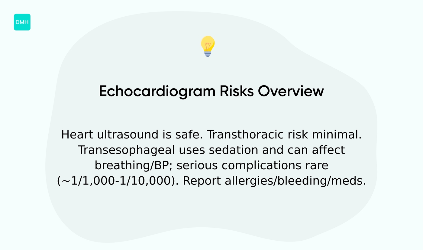
An echocardiogram is a heart ultrasound that uses sound waves. It doesn’t use ionizing radiation.
Transthoracic echocardiogram, the standard chest probe test, carries minimal risk. Patients may feel mild discomfort from the gel or pressure. Allergic reactions to gel or latex are uncommon.
Transesophageal echocardiogram uses a probe placed in the throat for clearer images. Sedation is often used. Sedation can cause breathing or blood pressure changes in some people.
Available research suggests serious complications such as esophageal injury occur in about 1 in 1,000 to 1 in 10,000 procedures. The exact rate can vary from person to person.
Stress echocardiogram evaluates the heart under exertion. Exercise or medication used to stress the heart can trigger chest pain or arrhythmia in a small number of cases.
Major adverse events appear to be uncommon, often on the order of 1 in several thousand tests according to clinical reports. Mild side effects may include throat soreness after transesophageal echo and temporary lightheadedness after stress testing.
Mention any allergies, bleeding disorders, swallowing problems, or current medications before the test to help clinicians plan care. Not a medical advice, content for educational purposes, consult a professional.
You might also like: AST (SGOT) In Blood Test: What It Is And Why It Matters
How accurate is an echocardiogram
An echocardiogram uses ultrasound to image heart structure and blood flow. Accuracy depends on the question it tries to answer.
Heart ultrasound accuracy for structural problems stays high. Transthoracic echocardiography detects severe valve disease and chamber enlargement with sensitivities near 85–95% in many studies.
Transesophageal echocardiogram sensitivity for detecting valve infections (infective endocarditis) often reaches about 90%, while transthoracic sensitivity can be near 50–60%.
Stress echocardiogram sensitivity for coronary artery disease averages about 75–85% sensitivity and 80–90% specificity in pooled analyses. Resting echo has low sensitivity for isolated coronary stenosis.
Ejection fraction estimates usually agree with cardiac MRI within roughly 5–10 percentage points. Limitations include operator skill, patient body habitus, and acoustic windows.
Small wall motion changes and mild coronary blockages can be missed. If blocked arteries are suspected, clinicians may request coronary CT angiography, nuclear perfusion testing, or invasive angiography via heart catheterization.
Cardiac MRI helps when tissue detail matters. Evidence indicates echocardiography reliably identifies structural heart disease. Detection of coronary blockages can vary from person to person and often requires additional tests.
For informational purposes only. Always consult a qualified healthcare professional for medical advice specific to your situation.
Read also: What Is Globulin In Blood Test: Complete Guide
How much does an echocardiogram cost
Patients often ask about echocardiogram cost. Prices depend on test type, location, and insurance status.
- Transthoracic echocardiogram: $200–$1,000 at outpatient clinics. Hospital outpatient billing may reach $500–$1,500.
- Transesophageal echocardiogram: $800–$3,000 because of sedation and specialized equipment.
- Stress echocardiogram: $400–$1,500 depending on exercise testing or medication stress.
- 3D echo and advanced Doppler: add $200–$800 for specialized imaging or post-processing.
Medicare and many private insurers cover medically necessary echocardiography. Coverage often depends on diagnosis codes and prior authorization. Copays and deductibles affect out-of-pocket cost.
Factors that change price include facility type, regional costs, provider fees, need for sedation, length of monitoring, and whether the test is inpatient or emergency. Out-of-network billing can increase expenses substantially.
Patients who face high bills can explore financial assistance options. Hospital financial aid, sliding scale clinics, Medicaid, charity care programs, and interest-free payment plans reduce immediate cost.
Asking the billing office for an itemized estimate helps plan ahead. Available research and coverage rules can vary from person to person. For informational purposes only. Always consult a qualified healthcare professional for medical advice specific to your situation.
You’ll also like: How to Understand Albumin in Blood Test Results
Educational notice: This content is provided for informational and educational purposes only and is not intended as medical advice. Always consult a qualified healthcare professional for medical concerns.