Basophils vs eosinophils highlights clear differences in appearance, abundance, and immune role.
Basophils drive early vascular changes and allergy responses.
Eosinophils specialize in attacking parasites and modulating tissue inflammation.
Basophils carry coarse dark purple-black granules that often obscure a lobed nucleus. They’re the largest granulocytes and make under 1% of circulating white blood cells.
Eosinophils show bright red-orange granules and a distinct bilobed nucleus. They usually account for 1 to 4% of white cells.
Basophils trigger inflammation by releasing histamine and heparin. They bind IgE and degranulate during allergic reactions, increasing blood flow to damaged tissue within minutes.
Eosinophils attack multicellular parasites by releasing toxic granule proteins. They release major basic protein that damages parasite membranes and disrupts ion balance.
Both cells are types of granulocytes. They degranulate to shape local immune responses, but their targets differ significantly.
IL-5 controls eosinophil production and survival and links to the Th2 response. Basophils assist allergic inflammation and aid early vascular changes through histamine release.
Normal basophil counts sit below 1% or below 0.3 x10⁹/L. Normal eosinophil counts range 1 to 4% or 0.0 to 0.5 x10⁹/L.
Basophilia points to chronic allergy, inflammation, or myeloproliferative disease. Eosinophilia often indicates parasitic infection, asthma, or allergic disease.
Recognizing these differences helps clinicians interpret differential white blood cell counts accurately. Use counts to flag basophilia or eosinophilia and guide further testing.
If you see abnormal values, discuss them with your clinician for targeted evaluation and management.
Basophils function in the immune system
Histamine and heparin release
Basophils act quickly at damaged tissue sites. They degranulate and release histamine and heparin from cytoplasmic granules.
Histamine dilates small blood vessels and increases vascular permeability. This boosts blood flow and lets neutrophils and plasma proteins reach injured tissue within minutes.
Heparin inhibits local clotting so immune cells move freely through the tissue.
Basophils function ties directly into allergic reactions via IgE-triggered degranulation and mediator release. For example, when someone with a peanut allergy encounters peanut protein, basophils degranulate rapidly and flood tissues with histamine.
Understanding these steps helps you interpret symptoms and lab findings in a basophils vs eosinophils context.
Knowledge of histamine release from basophils and heparin action guides choices like antihistamines and careful anticoagulant use in inflamed tissue.
IgE binding and degranulation
Basophils start allergic reactions by binding IgE and releasing granule contents. They represent under 1% of circulating leukocytes.
They display high-affinity FcεRI receptors that hold IgE on the cell surface.
Allergen molecules bridge IgE on neighboring FcεRI. This IgE cross-linking triggers a rapid activation cascade.
Calcium influx and kinase activation drive granule fusion with the membrane.
Cells eject histamine, heparin, leukotrienes, and cytokines like IL-4 and IL-13. That histamine release causes vasodilation and permeability within seconds to minutes.
Clinicians can detect activation by measuring CD63 on basophils.
This pathway separates basophils from eosinophils in allergic responses. Knowing this mechanism helps you interpret allergy tests and plan targeted therapy.
Basophils vs mast cells
Use basophils vs mast cells as a simple framework: location, circulation, and granule composition.
Basophils circulate in blood and make up under 1% of white cells. Mast cells lodge in skin, lungs, and gut tissue — they don’t circulate freely.
Basophil granules trigger histamine release and heparin after IgE cross-linking.
Mast cell granules contain similar mediators plus proteases and cytokines that persist in tissue. Basophils drive rapid systemic allergic signals and support early inflammation.
Mast cells sustain local inflammation and help remodel tissue over time.
Typical basophil counts run 0–300 cells/µL or major basic protein.
Clinicians gain value by checking both circulating basophils and tissue mast activity to explain persistent allergy symptoms. Use this view to guide your lab interpretation and care decisions.
Eosinophils function and role in immunity
How eosinophils fight parasites
Eosinophils target multicellular parasites quickly and directly. They make up about 1–4% of white blood cells.
They patrol tissues and respond to antibody-coated helminths.
They bind IgE or IgG on parasite surfaces and trigger degranulation. That releases major basic protein and toxic granule contents such as eosinophil cationic protein and eosinophil peroxidase.
These toxic proteins punch holes in parasite membranes, disrupt ion balance, and impair parasite metabolism. Parasite larvae often lose motility within hours and die after sustained exposure.
For instance, eosinophils attacking Schistosoma larvae coat the worm surface and release granule proteins that cause membrane damage visible under electron microscopy.
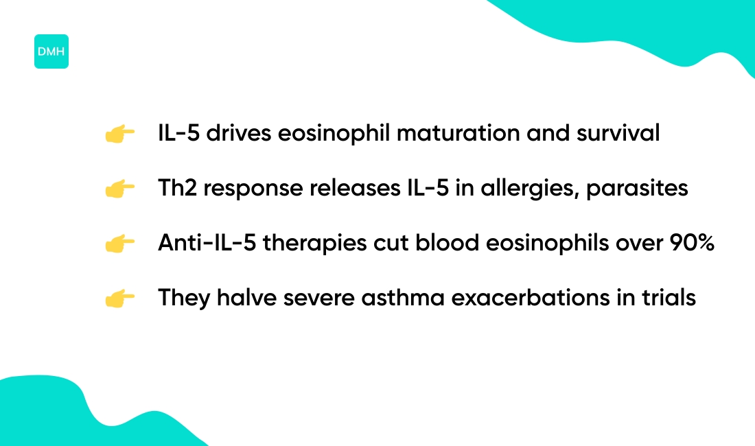
IL-5 increases eosinophil production and boosts this response during Th2-driven infections. Eosinophil degranulation is a powerful defense, though it can harm host tissue in conditions like asthma.
IL-5 and eosinophil regulation
Interleukin-5 steers bone marrow precursors into the eosinophil lineage. IL-5 drives eosinophils to mature, activate, and resist apoptosis.
IL-5 boosts eosinophil survival. Survival can rise from about 24 hours to several days in laboratory tests.
IL-5 triggers tissue recruitment and primes cells for degranulation.
Th2 immune response signaling releases IL-5 during allergic and parasitic reactions. Eosinophil degranulation releases major basic protein that damages parasites and harms tissue.
Targeting IL-5 cuts eosinophilia and improves symptoms.
IL-5 signals through the IL-5 receptor alpha on eosinophils. Therapies block IL-5 or its receptor to reduce counts.
Clinical trials show anti-IL-5 agents cut blood eosinophils by over 90%.
They reduce severe asthma exacerbations by about half in many trials. We recommend checking eosinophil counts if you have asthma or unexplained tissue inflammation.
A clinic visit and targeted testing can guide therapy choices.
Eosinophils in allergic reactions
Eosinophils drive allergic inflammation, worsen asthma, and damage tissue. In basophils vs eosinophils comparisons, eosinophils specialize in tissue infiltration and sustained damage.
They move into mucosa after Th2 signaling and IL-5 boosts their numbers. They release granule proteins including major basic protein, eosinophil peroxidase, leukotrienes, cytokines, and reactive oxygen species.
Degranulation injures airway epithelium and sustains bronchial hyperreactivity.
Eosinophils interact with mast cells, basophils, and T cells to amplify inflammation and recruit more immune cells. High eosinophil counts define eosinophilia and often appear in asthma and eosinophilic esophagitis.
Absolute counts above 500 cells/µL suggest significant involvement.
Measuring counts helps track response to anti-IL-5 therapies. We monitor eosinophils for treatment decisions.
Targeting IL-5 regulation cuts exacerbations in many patients.
What causes high eosinophils on a blood test
High eosinophil counts usually signal immune activation against parasites or allergies. Normal absolute eosinophil counts range from 0 to 500 cells/µL.
Common causes include:
- Parasitic worms (helminths)
- Allergic reactions and atopic disease
- Asthma
- Autoimmune disease
- Eosinophilic disorders like eosinophilic esophagitis
- Drug reactions
Parasitic infections trigger eosinophil activation. Major basic protein releases during degranulation to damage parasites.
Allergic conditions drive a Th2 response with IL-5 that raises eosinophil production and survival.
Specific disorders such as hypereosinophilic syndrome cause sustained, very high counts that need specialist care. We recommend prompt follow-up for persistent elevations or counts above 1,500 cells/µL.
| Eosinophil Level | Absolute Count | Clinical Action |
|---|---|---|
| Normal | 0–500 cells/µL | Routine monitoring |
| Mild elevation | 500–1,500 cells/µL | Review history, repeat test |
| Moderate–severe | >1,500 cells/µL | Specialist referral, targeted workup |
Tell your clinician about travel, pets, and recent medications. Order stool ova and parasite testing when exposure risk exists.
Request allergy testing or pulmonary evaluation for asthma symptoms.
Refer to a hematologist or immunologist for unexplained, persistent eosinophilia. Reviewing history and simple tests finds most causes quickly.
Check your complete blood count alongside notes on basophils to understand the full granulocyte picture.
What causes high basophils on a CBC
High basophils most often point to allergic or inflammatory processes, or to bone marrow disorders. Basophils normally make up 0–2% of white blood cells and typically sit below 0.2 x 10⁹/L.
An unusual basophil count rise above 2% or 0.2 x 10⁹/L deserves a clinical workup. Common causes include:
- Chronic allergic response
- Persistent infections
- Autoimmune inflammation
- Myeloproliferative neoplasms (e.g., chronic myeloid leukemia)
- Hypothyroidism
- Recovery phases after acute infection
Myeloproliferative neoplasms, such as chronic myeloid leukemia, can raise basophils sharply. Symptoms vary — you may feel itching, hives, or unexplained fatigue.
A peripheral smear can show basophil morphology. Molecular testing like BCR-ABL PCR helps confirm chronic myeloid leukemia.
Repeat testing within weeks reduces false alarms from lab variability.
We advise repeating the CBC, checking a differential, and testing inflammatory markers. Order targeted infection tests if counts remain high or symptoms persist.
See more causes and recommended next steps at what does high basophils mean. Get prompt clinical evaluation for persistent basophilia.
Read also: High White Blood Cell Count: Causes, Symptoms and more
How to interpret a differential white blood cell count
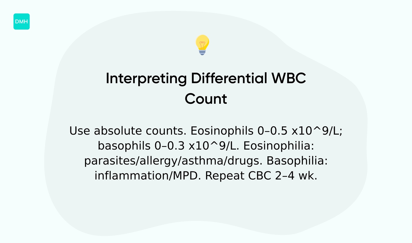
Read a differential white blood cell count to spot basophilia or eosinophilia quickly. We recommend using absolute counts over percentages for clearer decisions.
Normal ranges vary by lab. Eosinophils typically sit at 0–4% or 0–0.5 x10⁹/L (0–500 cells/µL).
Basophils usually measure 0–2% or 0–0.3 x10⁹/L (0–300 cells/µL).
One elevated percentage without a matching absolute rise misleads clinicians more often than not. Why does this matter? Because percentages shift when other cell types change, but absolute counts reflect true cell numbers.
High eosinophils, labeled eosinophilia, suggest parasites, allergic disease, asthma, drug reactions, or certain autoimmune conditions. High basophils, labeled basophilia, point toward chronic inflammation, myeloproliferative disorders, or persistent allergic states.
Follow simple checks to act on abnormal values:
- Confirm absolute count on repeat CBC within 2–4 weeks.
- Match results to symptoms like rash, wheeze, fever, or weight loss.
- Order targeted tests: stool O&P for parasites, serum IgE, CRP, or thyroid panel.
- Refer to hematology if absolute eosinophils exceed 0.5 x10⁹/L persistently or basophils exceed 0.3 x10⁹/L without clear cause.
Check the lab reference and detailed differential ranges on the CBC with differential values and meanings page for local cutoffs and method notes.
If abnormalities persist after two tests, get specialist evaluation to protect organs and guide therapy.
You’ll also like: Leukocytosis: definition, causes, symptoms and treatments
Basophilia and eosinophilia clinical significance
Treat abnormal basophil or eosinophil counts as signals to investigate underlying causes. They point to allergic reactions, parasitic infection, autoimmune disease, or blood disorders.
Start with a CBC with differential. Order repeat testing for mild, transient changes.
Consider bone marrow biopsy for marked, persistent elevations.
For details on basophil interpretation, consult what is basophils in blood test during your diagnostic review. Treatment links to the cause.
Use antihistamines and corticosteroids for allergy-driven counts. Use antiparasitic therapy for confirmed parasites.
Use hematology referral for suspected myeloproliferative disease.
Know numeric thresholds. Normal basophils run near 0–1% of WBC.
Normal eosinophils sit around 0–6% or 0–500/µL. Eosinophils above 500/µL suggest tissue involvement and warrant therapy.
Matching treatment to the likely cause reduces unnecessary tests and speeds recovery. Basophilia and eosinophilia guide specific workups.
If you see high eosinophil counts, check travel, allergy history, and medication list.
When results are unclear, repeat the test and get specialty input. We recommend documenting symptoms and recent exposures, then discussing persistent abnormalities with your clinician within two weeks.
You might also like: Monocytes: low, high, absolute count and normal range, causes